
Case 10: Plication of rectus diastasis 2 years after twin-birth
Theresia Skytte Eriksen1, Helle Sjøstrand and Ida Felbo Pold1
- 1Department of Plastic Surgery, Herlev and Gentofte Hospital, Denmark
Abstract
Keywords: Rectus Diastasis, Plication, Abdominoplasty, following pregnancy Authors: Theresia Skytte Eriksen, Helle Sjøstrand and Ida Felbo Pold. Institution: Department of Plastic Surgery, Herlev and Gentofte Hospital, Denmark Abstract 38-year-old woman underwent plication of symptomatic diastasis of the rectus abdominal muscles after twin-birth by caesarean section. The diastasis had a maximal width of 6,6 cm just above the umbilicus and her symptoms included core-instability, back-pain, and a feeling of being unprotected around her abdomen especially while handling her children. Patient medical history 38-year-old woman had after twin-birth developed symptomatic diastasis of the rectus abdominal muscles. She had no comorbidities. She had an older child of 6 years, and the twins were 2 years old. All children were delivered by uncomplicated caesarean section. The patient described symptoms as back-pain, nausea, core instability, discomfort during running, and pain when encountering pressure to the abdomen especially while handling the children. Physical rehabilitation with physiotherapists had not been successful and hard to complete do to discomfort and pain. Before and After Patient examination The patient was initially examined 15 months postpartum and at an additionally examination two years postpartum by the same surgeon. A preoperative CT-scan at 15 months postpartum, showed a rectus diastasis with the maximal width of 6,6 cm just above the umbilicus. The objective examination confirmed those findings, and a small umbillical hernia was detected. No change in symptoms and width of the diastasis was found between the two examinations. Pre-operative considerations Physical training alone has not been found to reduce the width of the diastasis [1]. Surgery was in one randomized controlled trial superior to physical training with regard to reduction of pain and increase abdominal muscle strength [2]. The swedish guidelines currently recommend that surgery is indic
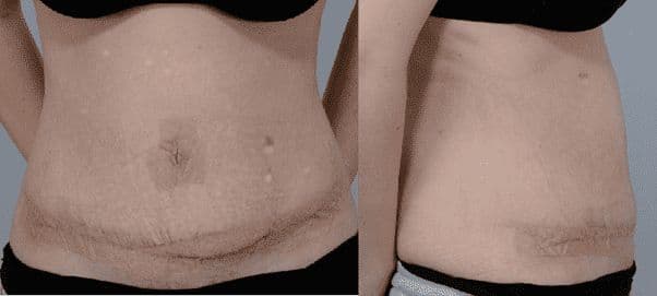
38-year-old woman had after twin-birth developed symptomatic diastasis of the rectus abdominal muscles. She had no comorbidities. She had an older child of 6 years, and the twins were 2 years old. All children were delivered by uncomplicated caesarean section. The patient described symptoms as back-pain, nausea, core instability, discomfort during running, and pain when encountering pressure to the abdomen especially while handling the children. Physical rehabilitation with physiotherapists had not been successful and hard to complete do to discomfort and pain.

The perioperative drawings are made in a standing position estimating the amount of loose skin expected to be excised.

Incision was made according to the caudal drawing and the skin was undermined at the plan of the fascia. The flap was raised up to 2-3 cm under the curvature of the ribs.
2

The maximum width was measured to 6,5 cm. The diastasis stretched from 1 cm below proc. xiphoideus to 4 cm above the symphysis.
3

The small umbilical hernia only contained fat which was excised and the defect in the fascia was closed with 3-0 Vicryl.
The plication of the diastasis was made with PDS 0 Loop-suture as a continuous suture. The suture was continued around the umbilicus. Starting cranially, locking the suture in the loop. At the end of the plication, one tread was cut and the two sutures were knotted together. The knot and suture ends were buried in the tissue.
4

Carefully measurement of the amount of extensive loose skin which can be surgically removed, so that the incision could be closed without too much tension. Approximately 550g of skin tissue was removed.
5

After thorough hemostasis the new hole for the umbilicus was made with a diamond-shaped incision. The umbilicus was sutured to the skin with 3-0 Novosyn and 5-0 Prolene.
The distal incision was closed in 3 layers with 2 layers of 3-0 Novosyn in the depth and the skin intracutaneously with 3-0 Biosyn.
Tranexamic acid were administrated both intravenously and in diluted state directly into the cavity.
A suction drain, and pain regulating catheter was placed. 8 ml of ropivacaine 7,5 mg/ml was administrated by the catheter at the end of the surgery. All incisions were covered by micropore tape.
The patient was discharged two days later.
6

The patient was seen two weeks postoperatively for suture removal around the umbilicus. There was no sign of infection, and no patient-reported pain. On the lower left side, a small seroma formation was detected.
7
How to cite this article
Theresia Skytte Eriksen, Helle Sjøstrand and Ida Felbo Pold. Case 10: Plication of rectus diastasis 2 years after twin-birth. Journal of Plastic, Breast & Reconstructive Surgery. 2025.
Open access. © 2025 The Author(s). Published by the Journal of Plastic, Breast & Reconstructive Surgery. Copyright to this case is shared between the author(s) and the journal; authors may reuse their own case for education, presentations and social media.
Keep reading
Related cases
Case 17: Immediate Proximal Row Carpectomy for combined Scaphoid and Lunate Dislocation Without Fracture
Jorge Jimenez Lopez, Anastasia Boptsi, Konstantinos Kalousis
Keywords: Hand surgery; wrist; carpectomy; scaphoid; lunate dislocation Authors: Jorge Jimenez Lopez, MD; Anastasia Boptsi, MD; Konstantinos Kalousis, MD Institution: Universitätsklinikum Schleswig-Holstein Lübeck Abstract Carpal dislocation fractures are uncommon injuries, most frequently associated with high-energy trauma. Combined carpal dislocations without associated bone fractures are even rarer. Delayed treatment can have serious consequences. The case presented here involves an 86-year-old man with dislocation of the scaphoid and lunate bones, as well as acute carpal tunnel syndrome resulting from the injury. PRC is mostly a salvage operation but given the severity of ligamentous disruption, the need to restore the patient's mobility and also the fact, that scaphoid was irreducible, a proximal row carpectomy (PRC) was performed to preserve wrist range of motion (ROM). Patient medical history The 86-year-old patient crashed the car into the wall of his garage at a speed of 30 km/h. As a result, the patient presented a dislocation of the lunate bone and the scaphoid. The patient lived alone and was completely independent. Regarding other conditions, he suffered from arterial hypertension and prostate cancer. Before and After Patient examination There was a visible deformity at the right wrist with bruising, and swelling. Additionally, a positive Hoffman-Tinel sign was observed with paresthesia in the median nerve area. A radiological examination of the hand was then performed, which revealed a dislocation of the scaphoid and lunate bones. No bone fractures were detected. Pre-operative considerations Given the severity of ligamentous disruption and the need to restore the patient’s mobility a proximal row carpectomy (PRC) was performed to preserve wrist range of motion (ROM). 1 Surgical Dorsal Exposure The surgical procedure involved exposing and splitting the 4th and 3rd extensor tendon compartments to facilitate access. The extensor pollicis longus (EPL) tend
Case 16: Utilizing the reverse radial forearm flap for burn wound management of the hypothenar part of hand
David Salim, MD, Msc
Keywords: Reverse radial forearm flap, Burn wound management, Hand surgery Authors: David Salim, MD and Taiba Alrasheed, MD, FRCSC, Msc. Institution: Department of Plastic- & Breast Surgery , Zealand University Hospital Roskilde, Denmark Abstract A 55-year-old man had sought and was admitted to the emergency department one week after sustaining a superficial second degree burn wound on the hypothenar region of the right hand after accidentally placing it on a hot stove. At first sight, the burn wound seemed quite superficial and conservative healing and bandaging was initially deemed sufficient with the aim of spontaneous healing. One week after the initial assessment in the emergency department, the patient was seen for a routine clinical control of the wound. At this consultation, the patient scored 4/4 positive Kanavel’s signs and flexor tenosynovitis was suspected. Afterwards immediate wound revision of the right hypothenar was conducted. During wound revision, considerable amounts pus was noted and furthermore the musculus abductor digiti minimi was avital and excised. Suitable wound dressing was applied, pus was sent for culture and sensitivity testing and the patient was gives appropriate peroral antibiotics. After numerous wound revisions and debridement, control of the infection at the orthopedic surgery department was achieved. The patient was then referred to the plastic surgery department for planning of soft tissue coverage with a reverse radial forearm flap. Patient medical history The patient was known to have substantial unregulated diabetes mellitus type two, but no other known medical comorbidities. Before and After Patient examination The patient was seen in an outpatient setting in the plastic surgery department for planning of surgery. The defect was located to the whole hypothenar region of the right hand. Substantial soft tissue loss was noted and the 3-5th flexor tendons were exposed. Pre-operative considerations A reverse radial forearm
Case 15: Closure of a complex defect after a dog bite wound of the middle third of the face
Dr. Guillermo Alvarez-Sanchez, Ignacio Lugo-Beltran
Keywords: Cheek reconstruction, face, cheek, forearm, free flap, Neurorrhaphy , radial forearm flap Authors: Dr. Guillermo Alvarez-Sanchez, MD ; Ignacio Lugo-Beltran, MD, Chief, Dr. Haro-Cruz Jorge Said, MD. Institution: Plastic and Reconstructive Surgery Division of Microsurgery, Hospital Angeles Metropolitano, Cuauhtémoc, Mexico. Abstract A 52 year old female who presents to the emergency room after receiving a dog bite in her right face, the defect is managed with a free radial forearm flap. Patient medical history 52 year old female, without past medical history, presented to the emergency room, referring she was attacked by her dog at her house, suffering a bite on her right cheek, with bleeding and loss of skin Before and After 1 Before photo - Arrival at the ER Facial asymmetry, with a complex wound on the right cheek, with skin loss, a defect approximately 10 x 9 cm, irregular borders, with muscle exposure, visualizing blood vessels and section of the buccal and zygomatic terminal nerve branches, active mild bleeding. Intact parotid gland and Stensen duct. Laceration of the zygomatic ligament. Difficulty mobilizing of the zygomatic and buccal branches muscle groups. Patient examination Facial asymmetry, with a complex wound on the right cheek, with skin loss, a defect approximately 10 x 9 cm, irregular borders, with muscle exposure, visualizing blood vessels and section of the buccal and zygomatic terminal nerve branches, active mild bleeding. Intact parotid gland and Stensen duct. Laceration of the zygomatic ligament. Difficulty mobilizing of the zygomatic and buccal branches muscle groups. Pre-operative considerations Given the circumstance of tissue loss, active bleeding and section of the nerve branches, also being a contaminated wound, we decided to go to the operating room immediately Due to tissue loss dimensions, we planned a radial free flap for tissue coverage 2 Debridement and assessing damages we cleansed, remodeled the irregular borders and clos