
Journal of Plastic, Breast & Reconstructive Surgery
Step-by-step surgical case reports, peer-reviewed by leading plastic surgeons and shared openly to help patients around the world.


Featured cases
Explore cases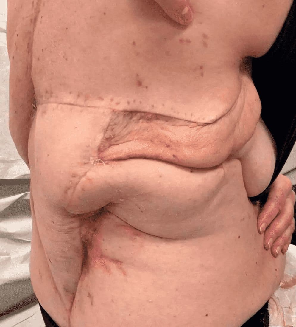
Case 1 - 2025: Lower back reconstruction with free ALT flap, free radial bridge flap and vein graft
Andrea Donfrancesco, Karolinska
Keywords: Double free flap, flap to flap, free radial bridge flap, radial conduit, vein graft, lower back defect. Authors: Andrea Donfrancesco, MD and Jenny Löfgren, MD, Karolinska University Hospital, Stockholm, Sweden Abstract A challenging case of midline mid-lower back defect is presented. The patient underwent previously vertebral chondrosarcoma resection, spine fixation and post-operative radiotherapy, and was re-operated 2 years after because of vertebral bone re-absorption and rupture of the previously done spinal fixation. The skin and soft tissue never healed and the patient had a 22 x 10 cm defect of the mid-lower back with exposed fixation material, exposed dura above the spinal cord, and radiotherapy damaged tissue around. No local reconstructive option was feasible. We reconstructed the defect with a free ALT, connected to a free radial bridge flap and a saphenous vein graft, hooked up to the thoracodorsal vessel, and a new spinal fixation was performed. Reconstruction was successful. Patient medical history The patient is previously healthy, has no past medical history. She developed a bulge on the back at T12 level. MRI and biopsy showed chondrosarkoma. She was operated at Karolinska University Hospital in May 2022 with sarkoma resection, spinal fixation T 10 to L3, primary skin closure, and post- operative radiotherapy. After 2 years the patient presented with osteoporotic reabsorption of bone, fusion/collapse of L1-L2, and rupture of a shaft of the previous fixation. The patient underwent again surgery by the spine surgeon at our hospital in July 2024 with extraction of the previously done fixation, and a new longer fixation was performed T5 to L5. Beacuse the soft tissues ware previously irradiated, a left pedicled muscle latissimus flap was performed to have better coverage of the upper potion of the defect. The wound ruptured and got infected, with expsure of the spinal fixation material; the patient received targeted IV antibiotics and the woun
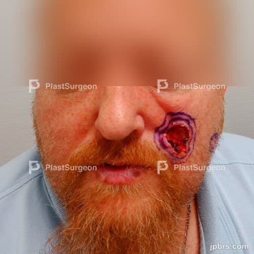
Case 1 (2023): Left Cheek Reconstruction with Combined VY- and Transposition Flap
Harald Welling, Michael Rose, Consultant, Roskilde Hospital, Denmark
50-year old male presented to the dept. of Plastic Surgery in 2022 with a larger eroding tumour on the left cheek. The tumour had been present for several years. Recently, a new tumour had also started to form on the lateral portion of the left cheek. Biopsi revealed basal cell carcinoma, nodular subtype for both carcinomas.
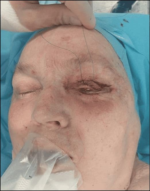
Case 02 2026: Tarsoconjuctival Flap for Eyelid Reconstruction
Ajla Sabitovic, Rikke Børthy Petersen, Nanja Gotland Sundstrup
Keywords: Basal Cell Carcinoma, Skin Cancer, Eyelid Reconstruction, Tarsoconjuctival flap. Authors: Ajla Sabitovic, MD, Rikke Børthy Petersen, MD, Nanja Gotland Sundstrup, MD. Abstract This case describes the use of a tarsoconjuctival flap for reconstruction of the lower eyelid following excision of a basal cell carcinoma in a 76-year-old woman. Due to the tumor size, depth and location near the limbus of the eye, direct closure was not possible. The tarsoconjuctival flap was chosen as the preferred reconstructive approach because of its ability to reconstruct all layers of the eyelid. Postoperative follow-up at six months demonstrated satisfactory aesthetic and functional outcomes. Patient medical history A 76-year-old woman presented with a biopsy-confirmed nodular basal cell carcinoma involving the right lower eyelid. Tumor excision was performed in general anesthesia with 3 mm margin and intraoperative frozen-section control, confirming tumor free margins. Resection resulted in a full-thickness defect involving approximately 90% of the lower eyelid. Reconstruction was performed with a tarsoconjuctival flap for the posterior lamella and a Tripier flap from the upper eyelid for the anterior lamella. Before and After Patient examination Clinical examination revealed a 12 x 10 mm elevated ulcerating lesion involving the right lower eyelid, extending into conjunctiva and limbus. Pre-operative considerations The patient presented with a full-thickness defect of the lower eyelid involving conjunctiva and the inferior eyelid margin. Due to the tumor size and location direct closure was not possible. Reconstruction with a flap was therefore required to secure reconstruction of all layers of the lower eyelid to give the eyelid full support and thereby secure its function. 1 Step 1 Preoperative skin markings outlining the tumor and 3 mm excision margin. 2 Step 2 Excision of the tumor was performed with a 3 mm margin, involving the conjunctiva, tarsal plate and lower eyelid
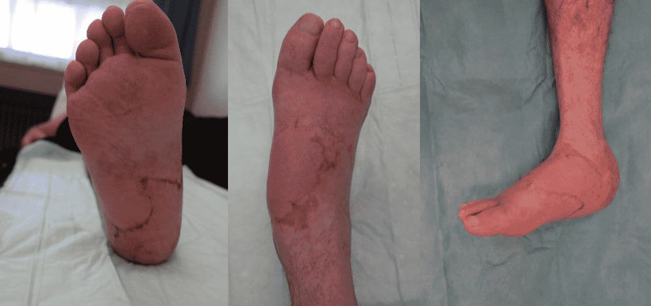
Case 2 - 2025: Reconstruction of an extensive foot defect using a combined sensory split latissimus dorsi and serratus anterior muscle flap
Dr. med. Luisa Lotter, PD
Keywords: Lower extremity; Flap; Reconstruction; Soft tissue; Trauma; Microsurgery; Foot; Latissimus Dorsi ; Authors: Dr. med. Luisa Lotter, Dr. med. Ilja Käch, PD Dr. med. Volker Schmidt. Institution: Kantonsspital St. Gallen, Switzerland Abstract A 67-year-old male patient sustained a complex foot injury with an open Lisfranc dislocation of the medial cuneiform bone and shaft fractures of the 2nd to 4th metatarsals in July 2024, as a result of a crush trauma. After initial osteosynthetic treatment, a pronounced necrosis developed in the area of the hindfoot and forefoot over time and an implant-associated wound infection. The patient was then transferred to our tertiary care hospital for specialized defect reconstruction. At first radical necrosectomy was performed, resulting in a 10 x 12 cm soft-tissue defect in the heel area, which extended medially and laterally. In the area of the forefoot, there was also a full-thickness wound measuring 4 x 3 cm with exposed osteosynthetic material, bone, and tendon. Patient medical history The patient has no pre-existing conditions. Blood tests on admission showed a blood glucose level and HbA1c value within the normal range. An angiography was performed, revealing a regular three-vessel supply to the affected lower right leg. Before and After Patient examination Upon admission to our hospital, on assessment, the 67-year-old male patient was in good general health with a slim nutritional status. On the heel area of the right foot was a 10 x 12 cm soft-tissue defect, which extended medially and laterally. In the area of the forefoot, there was also a full-thickness wound measuring 4 x 3 cm with exposed osteosynthetic material, bone, and tendon. Blood tests on admission showed a blood glucose level and HbA1c value within the normal range. An angiography was performed, revealing a regular three-vessel supply to the affected lower right leg. Pre-operative considerations The reconstruction of complex soft tissue injuries with exp
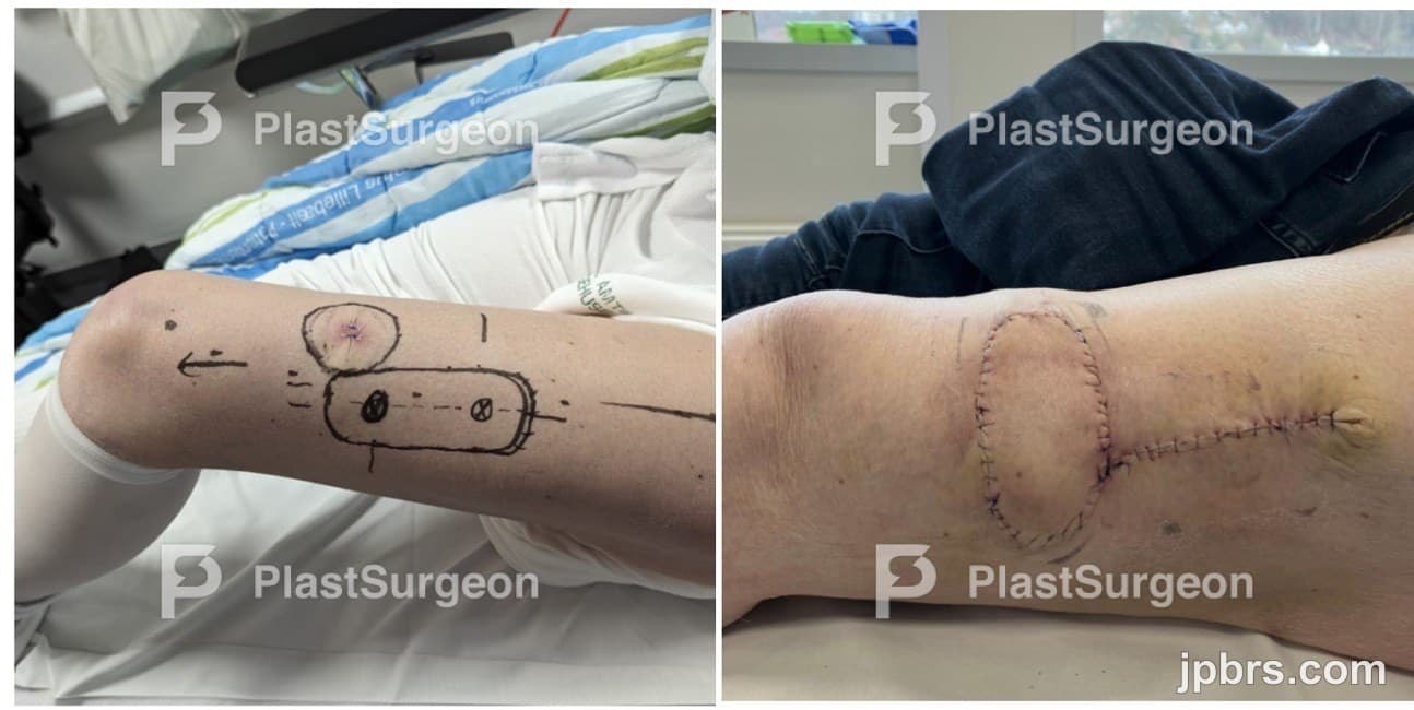
Case 2 (2024): Perforator-based soft tissue reconstruction around the knee, utilizing a D-POP ALT flap
Abdullah Najib Maalouf, Peter Sinkjær Kenney (MD, PhD)
A 73-year-old woman presented with malignant melanoma (pT2b-tumour) on her left thigh, located approximately 10 cm above the knee. The excision procedure aimed to remove the melanoma with a margin of 20 mm and perform sentinel node biopsy according to the protocols from Danish Melanoma Group (DMG). The patient had chronic obstructive lung disease, hypertension, hypercholesterolemia, ulcerative col
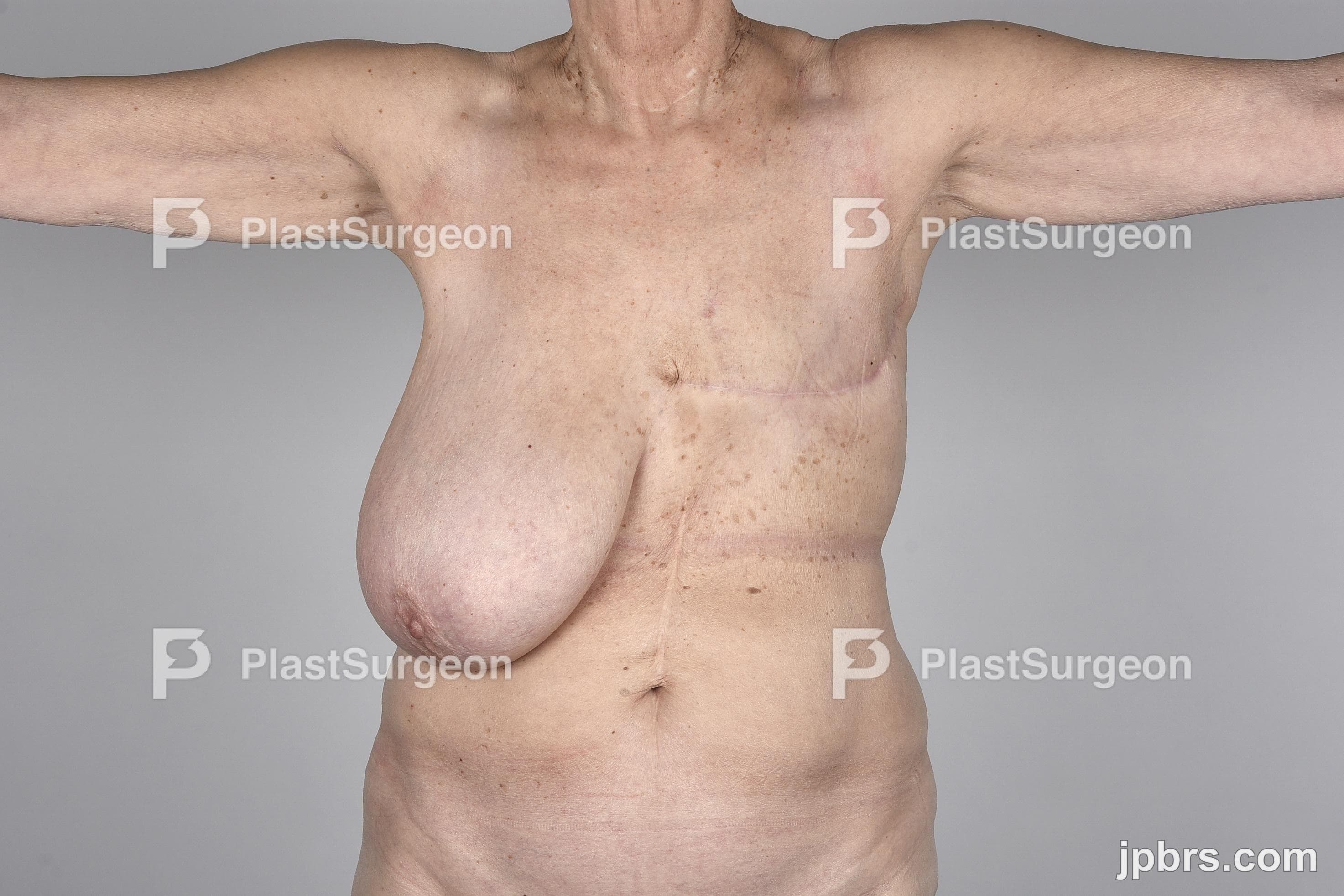
Case 2 (2023): Brystrekonstruktion med breast sharing-teknik
Nanja Gotland, Charlotte Uth, Alessandro Venzo, Rigshospitalet, Copenhagen, Denmark
En 62-årig kvinde blev henvist til sekundær brystrekonstruktion (SBR). Hun var tidligere mastektomeret pga. cancer mamma. Hun havde som barn nået den tilladte livstidsstråledosis pga. mb. Hodgkin og var derfor ikke egnet til en brystbevarende operation.
Most read
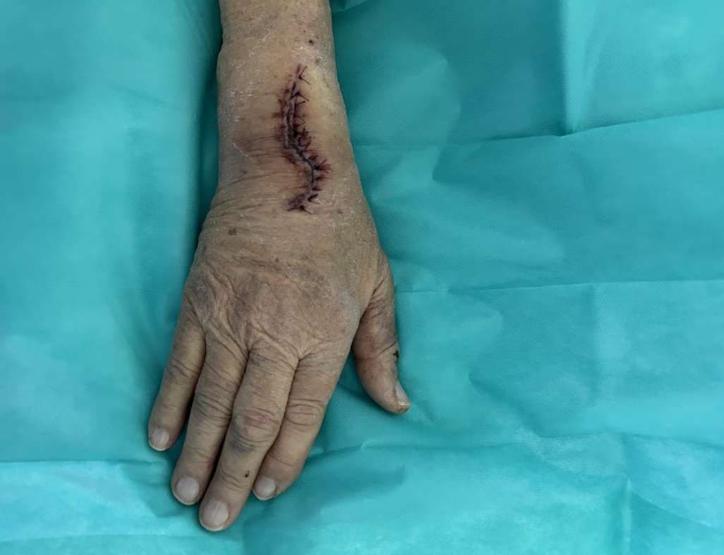
Case 17: Immediate Proximal Row Carpectomy for combined Scaphoid and Lunate Dislocation Without Fracture
Jorge Jimenez Lopez, Anastasia Boptsi, Konstantinos Kalousis
Keywords: Hand surgery; wrist; carpectomy; scaphoid; lunate dislocation Authors: Jorge Jimenez Lopez, MD; Anastasia Boptsi, MD; Konstantinos Kalousis, MD Institution: Universitätsklinikum Schleswig-Holstein Lübeck Abstract Carpal dislocation fractures are uncommon injuries, most frequently associated with high-energy trauma. Combined carpal dislocations without associated bone fractures are even rarer. Delayed treatment can have serious consequences. The case presented here involves an 86-year-old man with dislocation of the scaphoid and lunate bones, as well as acute carpal tunnel syndrome resulting from the injury. PRC is mostly a salvage operation but given the severity of ligamentous disruption, the need to restore the patient's mobility and also the fact, that scaphoid was irreducible, a proximal row carpectomy (PRC) was performed to preserve wrist range of motion (ROM). Patient medical history The 86-year-old patient crashed the car into the wall of his garage at a speed of 30 km/h. As a result, the patient presented a dislocation of the lunate bone and the scaphoid. The patient lived alone and was completely independent. Regarding other conditions, he suffered from arterial hypertension and prostate cancer. Before and After Patient examination There was a visible deformity at the right wrist with bruising, and swelling. Additionally, a positive Hoffman-Tinel sign was observed with paresthesia in the median nerve area. A radiological examination of the hand was then performed, which revealed a dislocation of the scaphoid and lunate bones. No bone fractures were detected. Pre-operative considerations Given the severity of ligamentous disruption and the need to restore the patient’s mobility a proximal row carpectomy (PRC) was performed to preserve wrist range of motion (ROM). 1 Surgical Dorsal Exposure The surgical procedure involved exposing and splitting the 4th and 3rd extensor tendon compartments to facilitate access. The extensor pollicis longus (EPL) tend
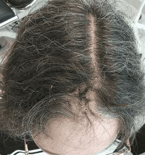
Case 20 2026: Staged Excision and Delayed Primary Closure of a large Nodular Basal Cell Carcinoma of the Anterior Scalp
Mikkel Halborg Sørensen, Nanja Gotland Sundstrup, Christian Lyngsaa Lang
Keywords: Basal cell carcinoma, scalp reconstruction, staged excision, primary closure, scalp biomechanics. Authors: Mikkel Halborg Sørensen, MD; Nanja Gotland Sundstrup, MD; Christian Lyngsaa Lang, MD. Institution: Department of Plastic Surgery and Burns, Rigshospitalet, Copenhagen University Hos-pital, Denmark Abstract A 48-year-old woman was referred with a non-radically excised nodular basal cell carcinoma of the anterior scalp complicated by wound infection and dehiscence. Re-excision with 3 mm margins to the galea was performed under local anesthesia. Given local inflammation and uncertain margin status, delayed reconstruction was planned pending histopathological confirmation. After clear margins were verified, controlled subgaleal undermining allowed tension-reduced primary closure. This staged strategy ensured oncologic safety while preserving hair-bearing scalp and avoiding unnecessary flap reconstruction. Patient medical history A 48-year-old woman was referred after incomplete excision of a nodular basal cell carcinoma (nBCC) of the anterior scalp performed in private plastic surgery practice. Histology demonstrated tumor involvement of the lateral margins, while the deep margin was free of tumor. Postoperatively, wound rupture and infection developed. Microbiology showed sparse growth of Staphylococcus aureus, and antibiotic therapy was initiated. The patient was otherwise non-smoking and healthy, with well-treated asthma, as her only relevant comorbidity. Before and After Patient examination Clinical examination revealed a 22 × 19 mm anterior scalp defect with limited perifocal erythema and minimal purulent discharge. No regional lymphadenopathy was palpable. Pre-operative considerations Initial histopathology demonstrated nodular basal cell carcinoma with focal lateral margin involvement and a clear deep margin, without perineural invasion or other high-risk features. In accordance with current recommendations for low-risk BCC, re-excision with 3 mm c
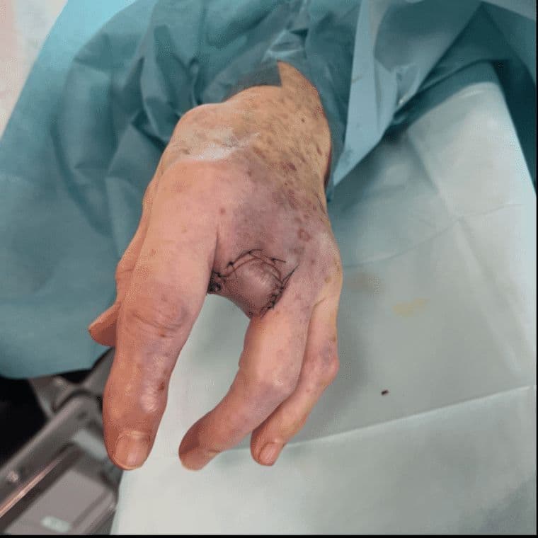
Case 23 2026: “Minimally Invasive Finger Amputation Using a Volar ‘Toilet seat’ Flap for Invasive Squamous Cell Carcinoma”
Claes Hannibal Killerich, Nikolaj Warming
Keywords: Squamous cell carcinoma; finger amputation; volar flap; local reconstruction; traction neurectomy; hand surgery. Authors: Claes Hannibal Killerich, Nikolaj Warming Abstract A 93-year-old woman presented with a chronic non-healing dorsal finger wound initially diagnosed as actinic keratosis. Repeat biopsy revealed invasive squamous cell carcinoma with joint and bone involvement. Due to the extent of invasion, distal amputation of the third finger was required. Reconstruction was performed using a volar “toilet seat” flap under digital nerve block to minimize surgical burden. The procedure achieved clear margins, preserved stump length, and avoided donor-site morbidity. This case highlights the importance of early re-biopsy of non-healing lesions and adapting surgical strategy to patient age, function, and comorbidity. Patient medical history A 93-year-old woman presented with a wound on the dorsal aspect of the proximal phalanx of the left third finger. A primary biopsy revealed actinic keratosis, and the lesion was treated several times with curettage and electrodesiccation by a private dermatologist. Despite repeated treatments under professional supervision, the wound failed to heal.Due to the persistent non-healing nature of the lesion, a repeat biopsy was performed, confirming the diagnosis of squamous cell carcinoma (SCC). The patient was referred to the Department of Plastic Surgery, Aalborg University Hospital, where a primary excision was performed with a 7 mm surgical margin. Histopathological examination demonstrated invasive tumor growth involving both joint and bone. The defect was subsequently reconstructed using a full-thickness skin graft. Before and After Patient examination The left third finger showed a healed full-thickness skin graft on the dorsal aspect of the proximal phalanx, covering the proximal interphalangeal (PIP) joint. There was no visible residual tumor, and no palpable lymphadenopathy in the left cubital fossa or axillary re
How it works
Submit
Authors submit a structured, step-by-step case with photos, pearls and references.
Peer review
Expert reviewers score each case across seven criteria and collaborate with the authors.
Publish
Approved cases are published open-access and indexed for the global surgical community.
Case Competition 2026 winners
The year's best step-by-step cases.
Top 3 International Cases 2026
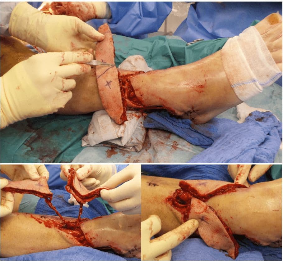
Case 19 2026: Split Perforator Flaps for bilateral ankle defects: A modern microsurgical approach to preserve function and reduce secondary thinning operations
Luisa Lotter¹, Marta Jezierska¹, Ilja W. Käch¹, Volker J. Schmidt¹ — Hospital St.Gallen, St.Gallen, Switzerland
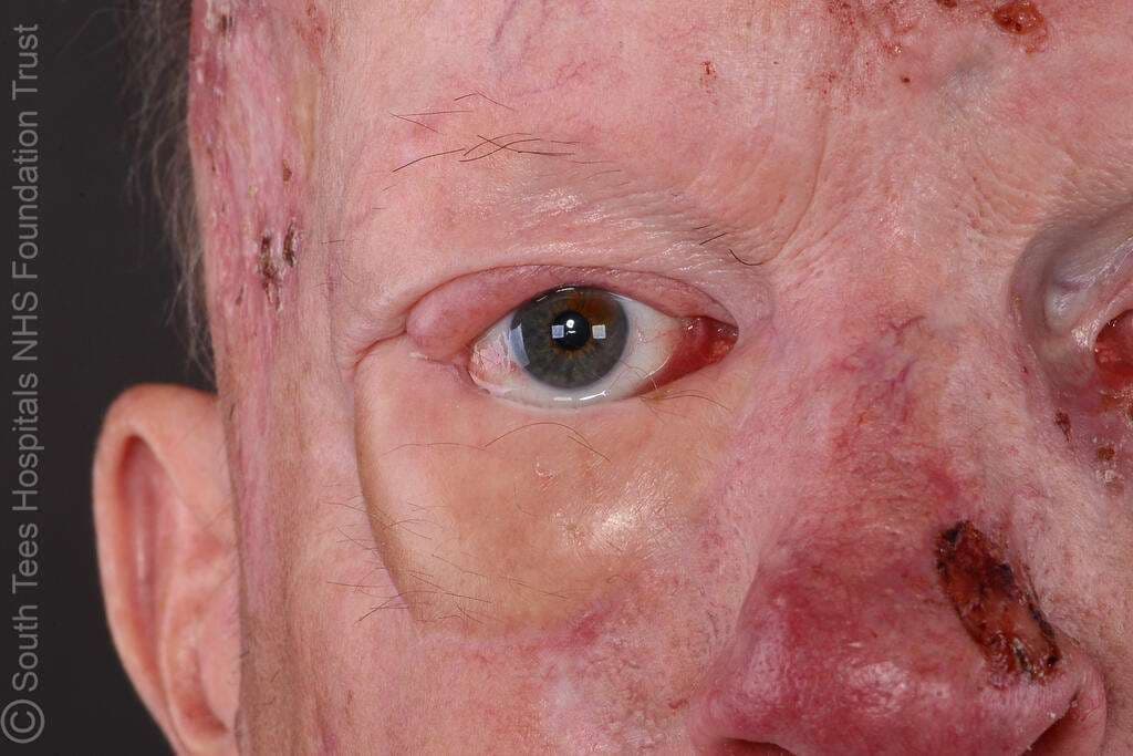
Case 21 2026: Reconstructing the Challenging Eyelid: Hard Palate Graft and Radial Forearm Free Flap Reconstruction in Gorlin–Goltz Syndrome
Miss Ketki Kaushal, Mr Oliver Bassett
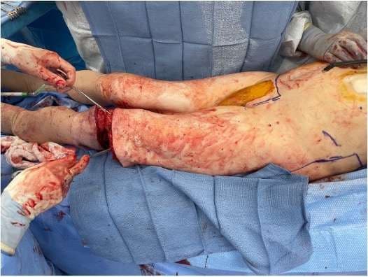
Case 13 2026: Fillet Flap Reconstruction Following Hemicorporectomy for Recurrent High-Grade Gluteal Sarcoma
Authors: Isha Joshi, Zachary Erlichman, Nia Nikkhahmanesh, Christina Chopra, Jason Ganz — Stony Brook University Hospital, Stony Brook, New York
Top 3 Danish Cases 2026
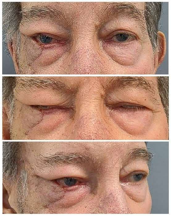
Case 08 2026: A Hella Lamella: A Three-Flap, Full-Thickness Lower Eyelid Reconstruction
Sondre F. Koren (MD), Jette B. Løvenwald (MD). Roskilde, SUH, Denmark
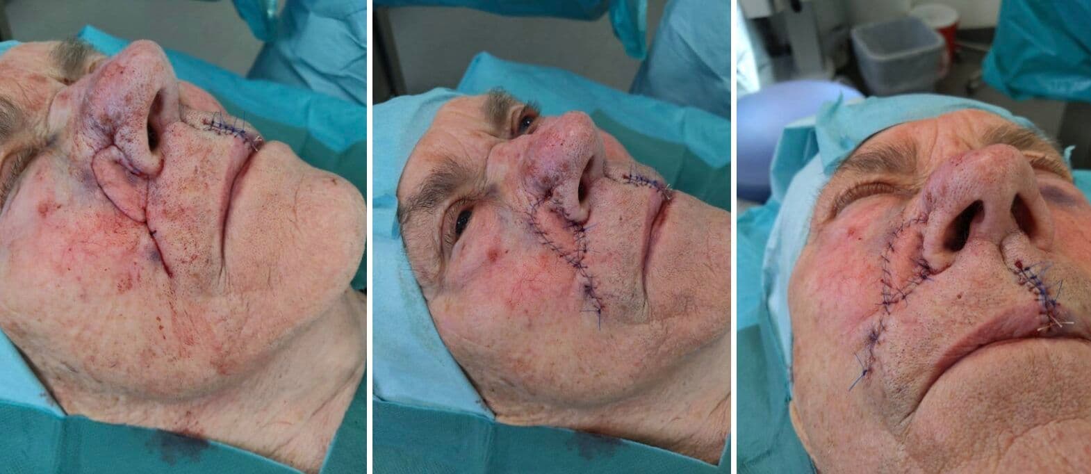
Case 12 2026: Closure of defect on ala nasi with modified shark island flap
Mickie Bang Christensen (MD), Magnus Avnstorp (MD) — Department of Plastic- & Breast Surgery, Zealand University Hospital Roskilde, Denmark
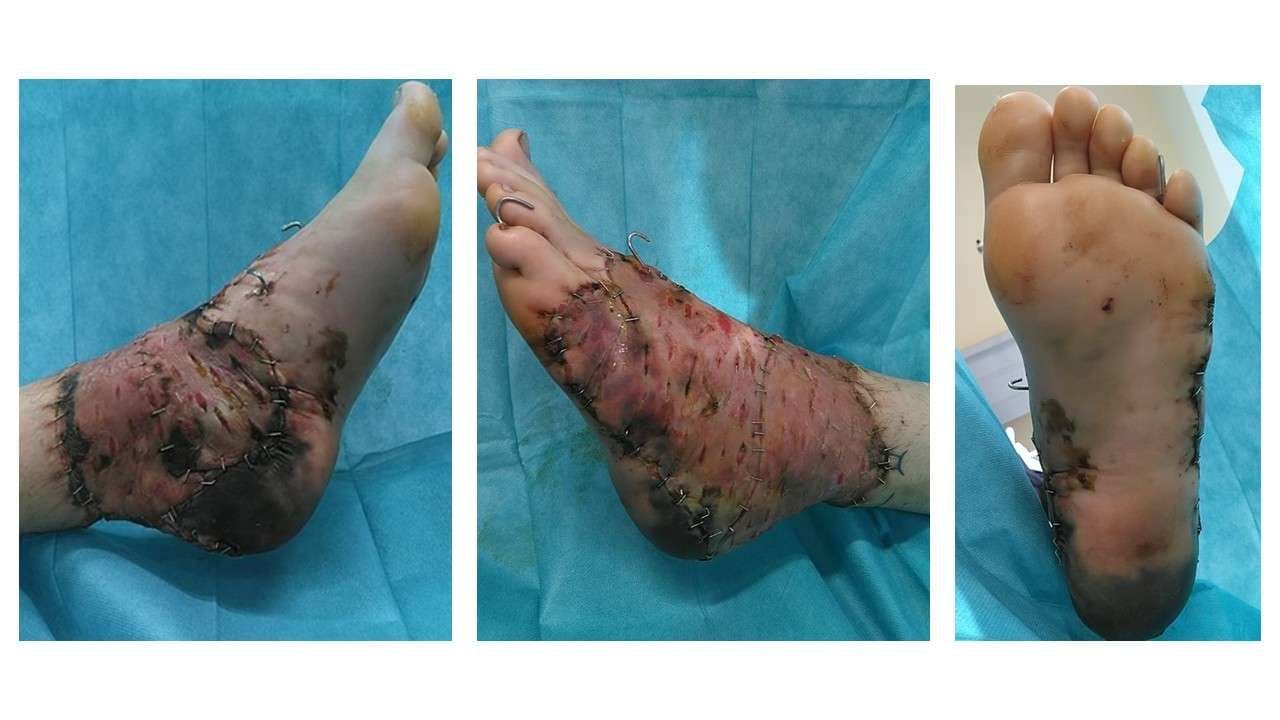
Case 16 2026: Severe Foot Degloving Injury Following a Forklift Crush: A Limb-Salvage Case
Cecilie Jansen, Emil Villiam Holm-Rasmussen, Mette Stueland Wolthers — University Hospital, Rigshospitalet, Denmark
Top 3 Trauma Cases 2026
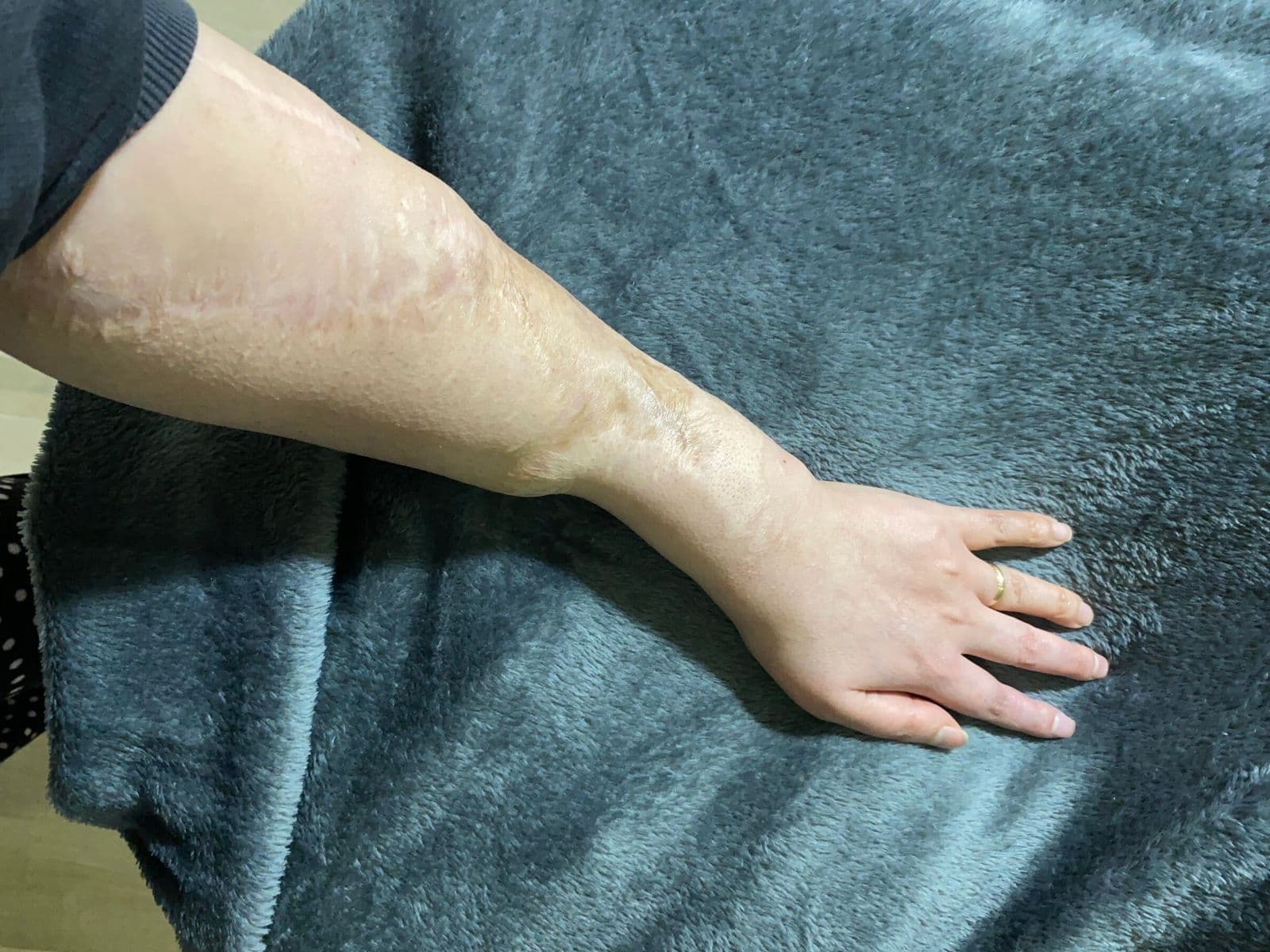
Case 03 2026: Emergency limb salvage with combined free vastus lateralis and anterolateral thigh flap after post-traumatic brachial artery graft thrombosis in a 16-year-old patient
Dr. Ibrahim Güler — Department of Plastic, Reconstructive and Aesthetic Surgery, Dokuz Eylül University, Izmir, Turkey; Department of Plastic, Aesthetic and Hand Surgery, Otto von Guericke University, Magdeburg, Germany
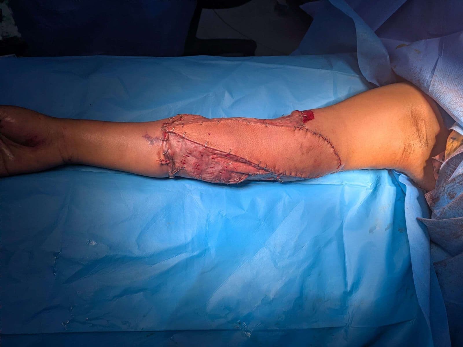
Case 24 2026: Ipsilateral Myocutaneous Latissimus Dorsi Flap for Traumatic Upper Limb Soft Tissue and Vascular Injury Following Road Traffic Accident
Sumaira Sattar — SMBB Institute of Trauma, Karachi, Pakistan
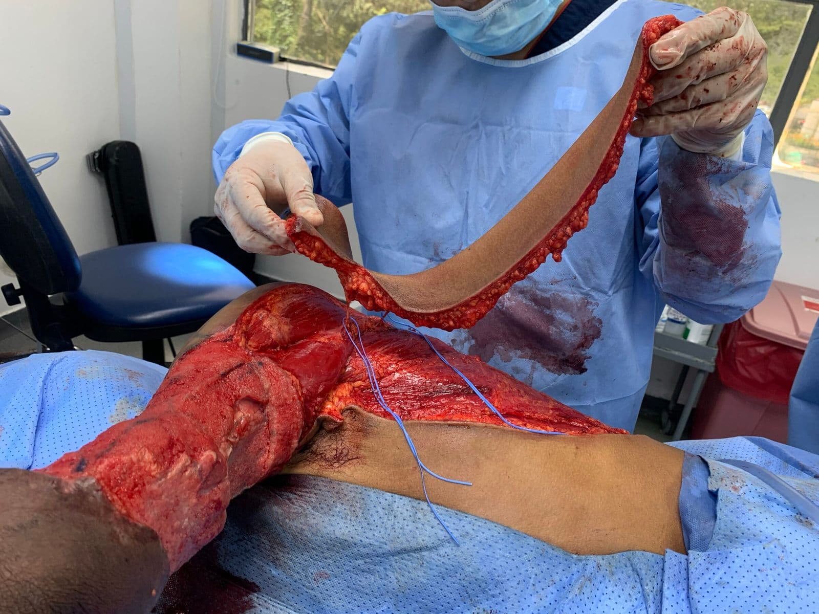
Case 10 2026: Propeller Flap Based on Two Perforators of the Scapular Artery for the Management of a Severely Traumatized Upper Extremity
Nelson Ramirez Lozano, Oncólogos de Occidente Manizales — Hospital de Santa Sofía Manizales, Colombia. Alejandro Angelillis Osorio. Surgical Assistant, Oncologos de Occidente, Manizales, Colombia
Have a case to share?
Join surgeons worldwide publishing peer-reviewed, open-access case reports — submission is straightforward and your draft saves as you go.