
Case 13 2026: Fillet Flap Reconstruction Following Hemicorporectomy for Recurrent High-Grade Gluteal Sarcoma
Authors: Isha Joshi1, Zachary Erlichman1, Nia Nikkhahmanesh1, Christina Chopra1, Jason Ganz1
- 1Stony Brook University Hospital, Stony Brook, New York
Abstract
Keywords: Hemicorporectomy, Fillet flap, Spare-parts surgery, Sarcoma reconstruction, Complex pelvic reconstruction Authors: Authors: Isha Joshi MD MBA, Zachary Erlichman MD, Nia Nikkhahmanesh MD, Christina Chopra MD, Jason Ganz MD Institution: Stony Brook University Hospital, Stony Brook, New York Abstract Background: Tissue from non-salvageable limbs may be used in reconstruction of complex defects and represent the principle of the “spare- parts” concept.[1] Fillet flaps advantages include soft tissue coverage of large defects, biocompatibility of an autologous donor, and donor tissue resembling native tissue due to proximity. Case: Fewer than 100 hemicorporectomies have been published in the literature.[2] We present the case of a 53-year-old male who underwent hemicorporectomy for recurrent gluteal sarcoma followed by reconstruction using a near-total thigh fillet flap. Conclusions: Fillet flaps provide soft-tissue coverage of large defects, biocompatible autologous tissue, and donor tissue resembling native anatomy. Patient medical history Primary Diagnosis: In 2015, the patient was diagnosed with a 15-cm high-grade (grade 3) spindle cell carcinoma thought to represent a fibrosarcoma with transformation to dermatofibrosarcoma protuberans, located superior to the buttock. The tumor was resected at an outside hospital (OSH) without perioperative complications. First Recurrence: In 2016, the patient presented to the OSH with recurrent mass in the tumor bed measuring 12.2 cm with erosion of lower coccyx, extension into posterior pelvis, and deviation of the anus. He underwent neoadjuvant radiation and systemic therapy. In 2017, he underwent tumor debulking and permanent diverting colostomy placement. After relocating to New York, he established care at our institution and subsequently underwent conversion of a loop colostomy to an end stoma with mucous fistula in 2020. Secondary Recurrence: In 2022, imaging demonstrated a recurrent tumor above the buttock. The pat
 BeforeAfter
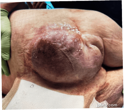
Primary Diagnosis: In 2015, the patient was diagnosed with a 15-cm high-grade (grade 3) spindle cell carcinoma thought to represent a fibrosarcoma with transformation to dermatofibrosarcoma protuberans, located superior to the buttock. The tumor was resected at an outside hospital (OSH) without perioperative complications. First Recurrence: In 2016, the patient presented to the OSH with recurrent mass in the tumor bed measuring 12.2 cm with erosion of lower coccyx, extension into posterior pelvis, and deviation of the anus. He underwent neoadjuvant radiation and systemic therapy. In 2017, he underwent tumor debulking and permanent diverting colostomy placement. After relocating to New York, he established care at our institution and subsequently underwent conversion of a loop colostomy to an end stoma with mucous fistula in 2020. Secondary Recurrence: In 2022, imaging demonstrated a recurrent tumor above the buttock. The patient underwent extensive oncologic resection including: abdominoperineal resection, sciatic nerve excision, and ischium excision by surgical oncology and orthopedic surgery. Plastic surgery reconstruction included a pedicled right vertical rectus abdominis muscle flap and a right vastus lateralis muscle flap. Following surgery, the patient's functional status progressively declined from ambulating with a cane to becoming bedbound. Between 2022 and 2024, he developed chronic urinary retention requiring indwelling foley catheter and progressive back pain requiring cordotomy and midline myelotomy Tertiary recurrence: In 2025, imaging demonstrated a large recurrent mass measuring 18 × 16 cm. After multidisciplinary evaluation, the patient elected to proceed with curative resection via hemicorporectomy. Plastic surgery was consulted for definitive soft-tissue reconstruction.

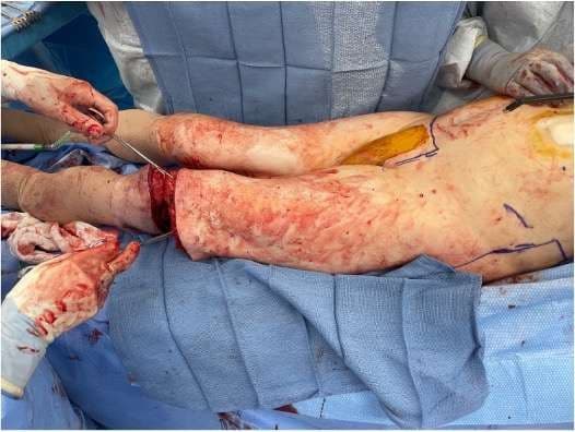
The flap was designed as a near-total fillet of thigh flap. The patient had a trochanteric and ischial pressure injury on his left leg posteriorly, which limited the use of the entire skin envelope, though the majority of the envelope was included with the flap. The longitudinal markings were made at the interval between the adductor magnus and biceps femoris while the lateral incision was made between the vastus lateralis and hamstrings.

A posterior to anterior approach was implemented with spinal surgery after marking the patient at the beginning of the case. Typically, after ligation of the inferior vena cava, Batson’s plexus becomes engorged, leading to a more challenging dissection and marked blood loss during division of the vertebral structures and spinal cord. Having experienced this problem in prior cases, a back-to-front approach was utilized with early division of the vertebral structures and spinal cord, pre-empting engorgement of Batson’s plexus, thus minimizing blood loss and neurogenic hypotension.

An incision was made just superior to the patella. After division of the quadriceps tendon, dissection proceeded distally to the femur. Medial dissection followed the interval between the adductor magnus and biceps femoris until the medial and lateral incisions were connected. The femoral artery entering the popliteal fossa was identified and controlled. The popliteal vessels were suture-ligated prior to transection.
3

With the vascular pedicle identified, the flap was elevated while incorporating the adductor musculature for additional coverage. The muscle was dissected off the femur from all directions as the flap was elevated in the distal to proximal direction. Care was taken to maintain the integrity of the quadriceps muscles as well as the gracilis, sartorius, adductor longus and their vascular pedicles. The flap was then elevated up to the level of the iliac crest.
4

Orthopedic surgery completed the pelvic disarticulation. A midline abdominal incision was performed, and the common iliac vessels were transected using a vascular stapler. The tumor was dissected from the external iliac vessels, and the internal iliac artery was divided.
5

The remaining posterior structures including psoas muscle, paraspinal muscles, and posterior soft tissue were divided, completing the hemicorporectomy specimen.
6
Early venous congestion of the flap was noted.
7

Leech therapy was used for approximately 5 days postoperatively when the flap was noted to be hyperemic and slightly tense. The team used a low threshold in deciding to initiate leech therapy given the significant change in habitus and somewhat unavoidable pressure to the flap. Gradual improvement of the hyperemia was noted over the course of the 5 days of leech therapy, with leeches aborted 24 hours after skin stability was noted.
8

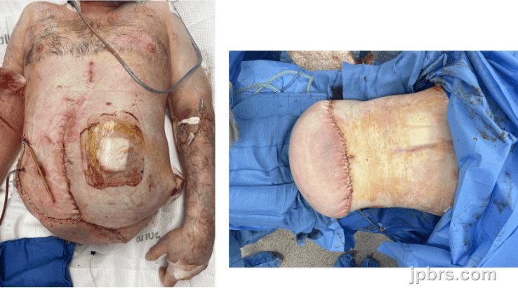
Patient underwent minor serial debridements intraoperatively to heal superficial dehiscences at the flap edges. Debridements were managed with wound vacs until good wound edge approximation was observed, at which point dressings were changed to mepilex. During this time the patient was downgraded from the Surgical ICU to the step down unit, transitioned to a pressure-offloading air mattress, and initiated physical therapy with a bedside trapeze to improve independence and activities of daily living.
9
The patient was medically optimized by the multidisciplinary team over the subsequent month and was discharged home with home health care at approximately 2 months postoperatively. At this point the patient was functional with most activities of daily living, including using a wheelchair. He was subsequently followed in the plastic surgery clinic on an outpatient basis with continued wound care to his fillet flap and no major sequelae to date.
10
- Strict bedrest
- Pressure offloading using air-fluidized mattress
- Trapeze for upper-body repositioning
- Physical therapy for activities of daily living
- Bedside psychotherapy offered
- Labs discontinued as soon as normalized
- Close monitoring for wound dehiscence
How to cite this article
Authors: Isha Joshi, Zachary Erlichman, Nia Nikkhahmanesh, Christina Chopra, Jason Ganz. Case 13 2026: Fillet Flap Reconstruction Following Hemicorporectomy for Recurrent High-Grade Gluteal Sarcoma. Journal of Plastic, Breast & Reconstructive Surgery. 2026.
Open access. © 2026 The Author(s). Published by the Journal of Plastic, Breast & Reconstructive Surgery. Copyright to this case is shared between the author(s) and the journal; authors may reuse their own case for education, presentations and social media.
Keep reading
Related cases
Case 20 2026: Staged Excision and Delayed Primary Closure of a large Nodular Basal Cell Carcinoma of the Anterior Scalp
Mikkel Halborg Sørensen, Nanja Gotland Sundstrup, Christian Lyngsaa Lang
Keywords: Basal cell carcinoma, scalp reconstruction, staged excision, primary closure, scalp biomechanics. Authors: Mikkel Halborg Sørensen, MD; Nanja Gotland Sundstrup, MD; Christian Lyngsaa Lang, MD. Institution: Department of Plastic Surgery and Burns, Rigshospitalet, Copenhagen University Hos-pital, Denmark Abstract A 48-year-old woman was referred with a non-radically excised nodular basal cell carcinoma of the anterior scalp complicated by wound infection and dehiscence. Re-excision with 3 mm margins to the galea was performed under local anesthesia. Given local inflammation and uncertain margin status, delayed reconstruction was planned pending histopathological confirmation. After clear margins were verified, controlled subgaleal undermining allowed tension-reduced primary closure. This staged strategy ensured oncologic safety while preserving hair-bearing scalp and avoiding unnecessary flap reconstruction. Patient medical history A 48-year-old woman was referred after incomplete excision of a nodular basal cell carcinoma (nBCC) of the anterior scalp performed in private plastic surgery practice. Histology demonstrated tumor involvement of the lateral margins, while the deep margin was free of tumor. Postoperatively, wound rupture and infection developed. Microbiology showed sparse growth of Staphylococcus aureus, and antibiotic therapy was initiated. The patient was otherwise non-smoking and healthy, with well-treated asthma, as her only relevant comorbidity. Before and After Patient examination Clinical examination revealed a 22 × 19 mm anterior scalp defect with limited perifocal erythema and minimal purulent discharge. No regional lymphadenopathy was palpable. Pre-operative considerations Initial histopathology demonstrated nodular basal cell carcinoma with focal lateral margin involvement and a clear deep margin, without perineural invasion or other high-risk features. In accordance with current recommendations for low-risk BCC, re-excision with 3 mm c
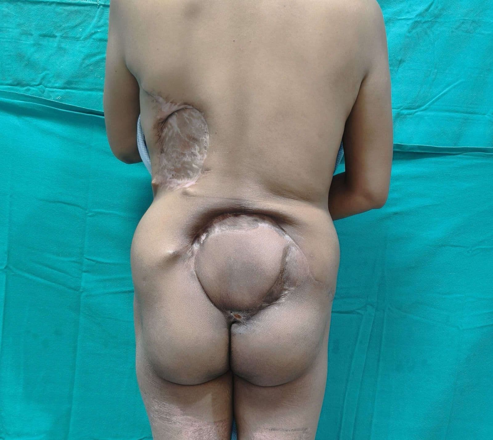
Case 22 2026: Superior Gluteal vessel: A rarely used recipient for free flap reconstruction of lumbosacral defects: a case report.
Tushar Dutta, Arun PS, Swati Sattavan
Keywords: Superior gluteal vessel, free flap reconstruction, lumbosacral defects Authors: 1. Tushar Dutta, Consultant Plastic Surgeon; 2. Arun PS, Consultant Surgical Oncologist; 3. Swati Sattavan, Consultant Surgical Oncologist; Institute: State Cancer Institute, Guwahati, Assam, India Abstract Lumbosacral defects that are too large for local flap reconstruction should be considered for microvascular free tissue transfer. However, lack of suitable recipient vessels in the area makes the task technically difficult. Options such as thoracodorsal and inferior gluteal vessels extended with vein grafts and AV loops have been described. Each option has its own drawbacks. Despite having a short course at its origin, the superior gluteal vessel is a viable alternative. We describe here a case where the vessel was dissected out with adequate length and used as a recipient for free flap reconstruction. Patient medical history The patient was a 41-year-old lady with no known comorbidities. She presented to us with a large lumbosacral mass that had been growing for one year. Before visiting our center, she had been operated twice at other institutions and had recurrences after each occasion. During the last three months the mass had started to grow rapidly and caused ulceration. There was no pain or fever. There was no neurological involvement of lower limbs. Patient had an ECOG status 1. Before and After Patient examination A 17x 22 cm mass was noted encompassing the left part of lumbosacral region and crossing the midline to the opposite side. The superior limit of the lesion was at the L3 vertebra and inferior extent was near the coccyx. Skin ulceration was noted at the center of the mass. On palpation, the mass was warm to touch, hard in consistency and immobile. Skin was fixed and fixity was also noted to the underlying muscle. No tenderness was elicited. Surrounding possible donor sites for flap harvest showed no scars. Pre-operative considerations Preoperative biopsy co
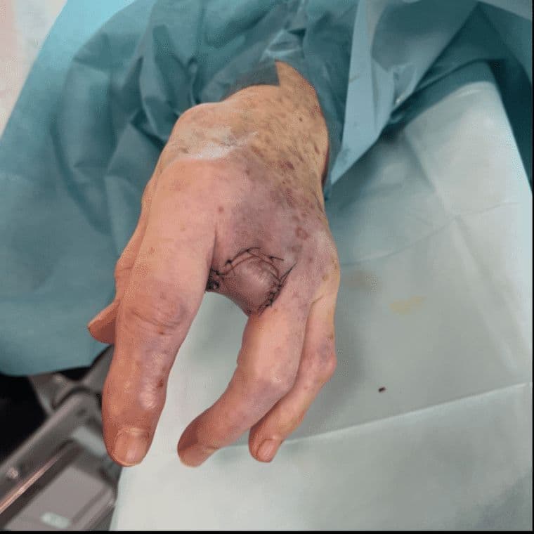
Case 23 2026: “Minimally Invasive Finger Amputation Using a Volar ‘Toilet seat’ Flap for Invasive Squamous Cell Carcinoma”
Claes Hannibal Killerich, Nikolaj Warming
Keywords: Squamous cell carcinoma; finger amputation; volar flap; local reconstruction; traction neurectomy; hand surgery. Authors: Claes Hannibal Killerich, Nikolaj Warming Abstract A 93-year-old woman presented with a chronic non-healing dorsal finger wound initially diagnosed as actinic keratosis. Repeat biopsy revealed invasive squamous cell carcinoma with joint and bone involvement. Due to the extent of invasion, distal amputation of the third finger was required. Reconstruction was performed using a volar “toilet seat” flap under digital nerve block to minimize surgical burden. The procedure achieved clear margins, preserved stump length, and avoided donor-site morbidity. This case highlights the importance of early re-biopsy of non-healing lesions and adapting surgical strategy to patient age, function, and comorbidity. Patient medical history A 93-year-old woman presented with a wound on the dorsal aspect of the proximal phalanx of the left third finger. A primary biopsy revealed actinic keratosis, and the lesion was treated several times with curettage and electrodesiccation by a private dermatologist. Despite repeated treatments under professional supervision, the wound failed to heal.Due to the persistent non-healing nature of the lesion, a repeat biopsy was performed, confirming the diagnosis of squamous cell carcinoma (SCC). The patient was referred to the Department of Plastic Surgery, Aalborg University Hospital, where a primary excision was performed with a 7 mm surgical margin. Histopathological examination demonstrated invasive tumor growth involving both joint and bone. The defect was subsequently reconstructed using a full-thickness skin graft. Before and After Patient examination The left third finger showed a healed full-thickness skin graft on the dorsal aspect of the proximal phalanx, covering the proximal interphalangeal (PIP) joint. There was no visible residual tumor, and no palpable lymphadenopathy in the left cubital fossa or axillary re