Case 23 2026: “Minimally Invasive Finger Amputation Using a Volar ‘Toilet seat’ Flap for Invasive Squamous Cell Carcinoma”
Claes Hannibal Killerich, Nikolaj Warming
Published · May 28, 2026
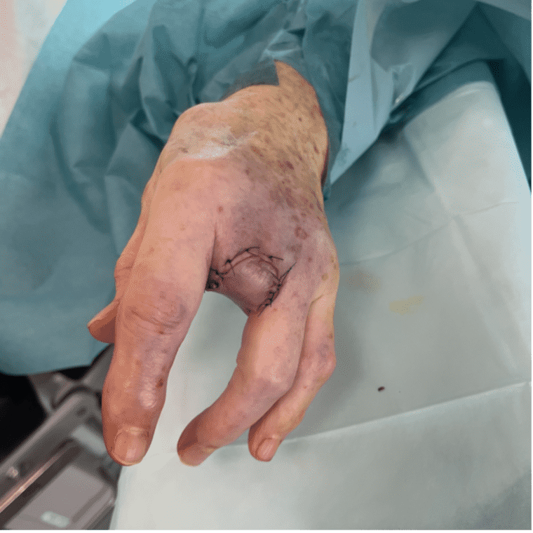
Keywords: Squamous cell carcinoma; finger amputation; volar flap; local reconstruction; traction neurectomy; hand surgery. Authors: Claes Hannibal Killerich, Nikolaj Warming Abstract A 93-year-old woman presented with a chronic non-healing dorsal finger wound initially diagnosed as actinic keratosis. Repeat biopsy revealed invasive squamous cell carcinoma with joint and bone involvement. Due to the extent of invasion, distal amputation of the third finger was required. Reconstruction was performed using a volar “toilet seat” flap under digital nerve block to minimize surgical burden. The procedure achieved clear margins, preserved stump length, and avoided donor-site morbidity. This case highlights the importance of early re-biopsy of non-healing lesions and adapting surgical strategy to patient age, function, and comorbidity. Patient medical history A 93-year-old woman presented with a wound on the dorsal aspect of the proximal phalanx of the left third finger. A primary biopsy revealed actinic keratosis, and the lesion was treated several times with curettage and electrodesiccation by a private dermatologist. Despite repeated treatments under professional supervision, the wound failed to heal.Due to the persistent non-healing nature of the lesion, a repeat biopsy was performed, confirming the diagnosis of squamous cell carcinoma (SCC). The patient was referred to the Department of Plastic Surgery, Aalborg University Hospital, where a primary excision was performed with a 7 mm surgical margin. Histopathological examination demonstrated invasive tumor growth involving both joint and bone. The defect was subsequently reconstructed using a full-thickness skin graft. Before and After Patient examination The left third finger showed a healed full-thickness skin graft on the dorsal aspect of the proximal phalanx, covering the proximal interphalangeal (PIP) joint. There was no visible residual tumor, and no palpable lymphadenopathy in the left cubital fossa or axillary re
References
- Reconstruction After Wide Excision of Primary Cutaneous Melanomas: Part II–the Extremities. Moncrieff MD, Thompson JF, Quinn MJ, Stretch JR. The Lancet. Oncology. 2009;10(8):810-5. doi:10.1016/S1470-2045(09)70121-4.
- Treatment of tendons in finger amputations and description of a new instrument. Webster, George V. Surgery, Volume 17, Issue 1, 102 – 108
- Scott BB, Winograd JM, Redmond RW. Surgical Approaches for Prevention of Neuroma at Time of Peripheral Nerve Injury. Front Surg. 2022 Jun 27;9:819608. doi: 10.3389/fsurg.2022.819608. PMID: 35832494; PMCID: PMC9271873.
- Calderazzi F, Menozzi M, Nosenzo A, Galavotti C, Pogliacomi F, Ceccarelli F. Single ray amputation in traumatic injury of the hand: review of literature. Acta Biomed. 2018 Oct 1;90(1-S):14-23. doi: 10.23750/abm.v90i1-S.7677. PMID: 30714994; PMCID: PMC6503410.