
Case 25 2026: Closed Reduction of a Distal Radius Fracture in the Emergency Department
Maria Lerche Mortensen and Philip Månsson1
- 1Department of Orthopedic Surgery, SUH Nykøbing Falster, Denmark
Abstract
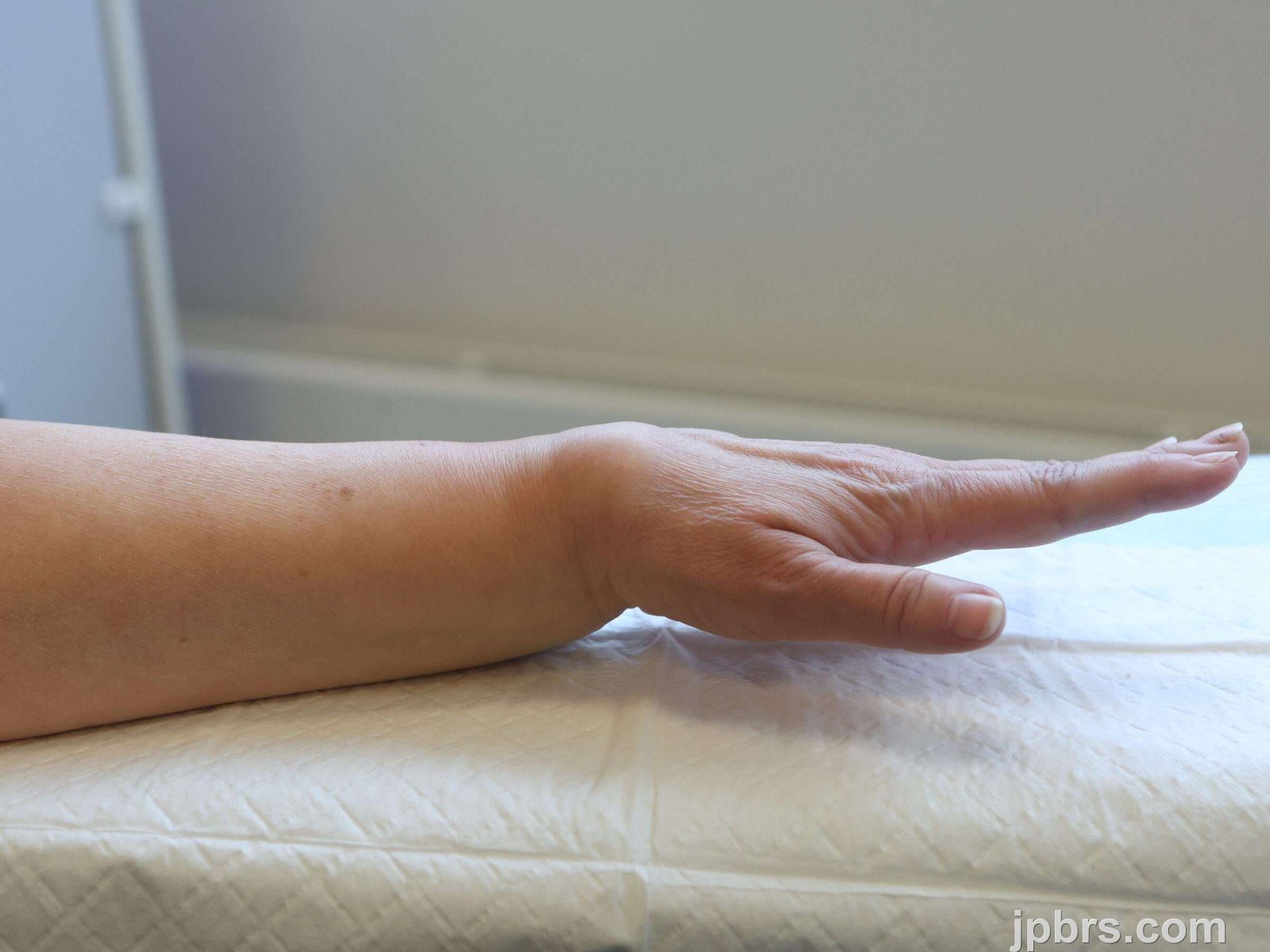
Keywords: Distal radius fracture, closed reduction, fracture management, Colles’ fracture, orthopedic injury Authors: Maria Lerche Mortensen and Philip Månsson. Institution: Department of Orthopedic Surgery, SUH Nykøbing Falster, Denmark Abstract A 62-year-old female presented to the emergency department after a fall from standing position, attempting to break the fall with her left hand. She presented with pain, swelling, and a visible deformity of the wrist. A distal radius fracture of Colles’ type was diagnosed. She was treated in the emergency department in accordance with local guidelines, with closed reduction using both Chinese finger traps and manual reduction with a hematoma block for analgesia. Finally, a dorsal below-elbow cast was applied, and the patient was referred for follow-up after 10-12 days. Patient medical history This patient was active and employed, able to take care of herself, her family and household. She had no past fractures, no relevant medication, and no known allergies. Before and After Patient examination Patient history was obtained, involving circumstances of the trauma, including reasons to suspect underlying disease or other injuries. She fell while walking on a slippery surface, described the trauma as “just bad luck”, she reported no other complaints of pain, had intact memory, and had no cardiac symptoms prior to fall. The clinical examination included inspection, palpation, and function. Inspection: A bayonet deformity was observed, with significant swelling over the radial styloid. There were no wounds or discoloration. Palpation: There was a palpable step-off, and pain over the distal radius. Elbow, hand and fingers were palpated without pain. Therefore, there was no suspicion of other injuries. Function: Neurological and vascular integrity were intact. The motor function was tested: n. ulnaris – finger abduction with intact strength; n. radialis – extension of the MCP-joints; n. medianus – opposition of the first digit. The
 BeforeAfter
This patient was active and employed, able to take care of herself, her family and household. She had no past fractures, no relevant medication, and no known allergies.

Before the procedure, several features on the radiographs were assessed.
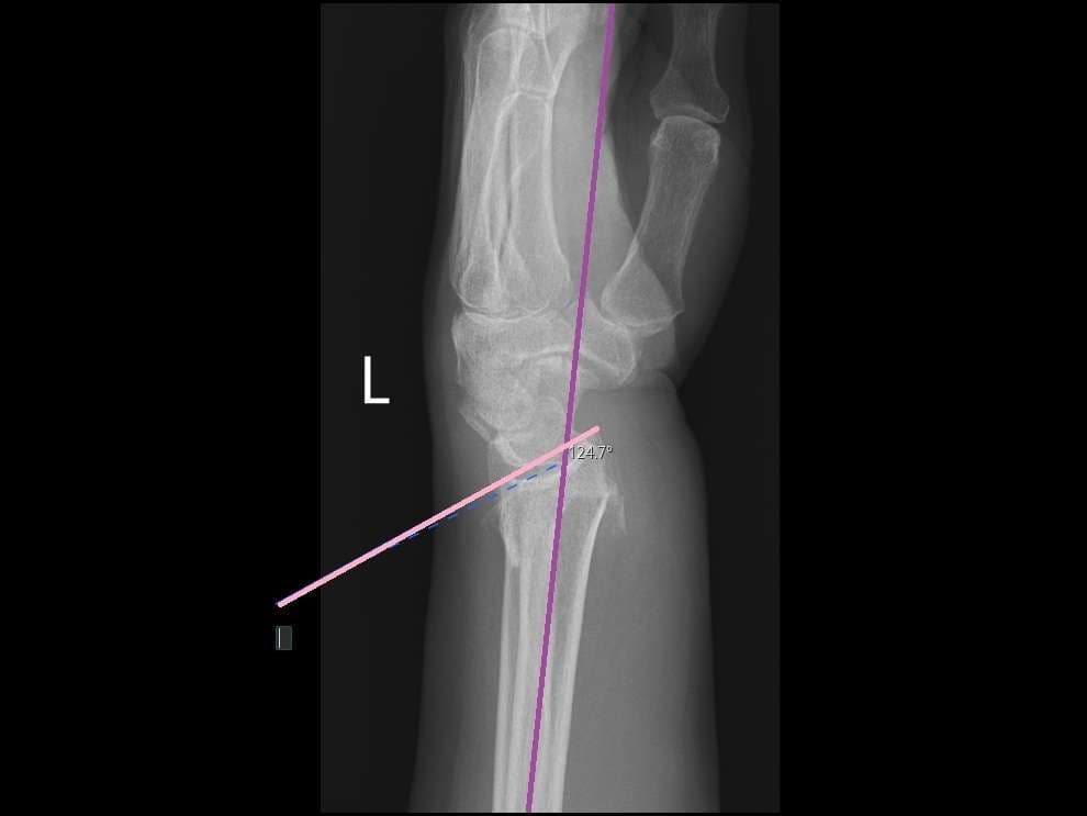
The degree of dorsal angulation was measured on the lateral view. It is defined as the angle between a line representing the long axis of the radial shaft (purple line) and a line representing the joint surface (pink line), intersecting the volar and the dorsal edges of the joint surface. The normal angulation is approximately 10 degrees volar, with minor anatomical variations. In this case, the dorsal angulation was 34 degrees dorsal, which was considered unacceptable.

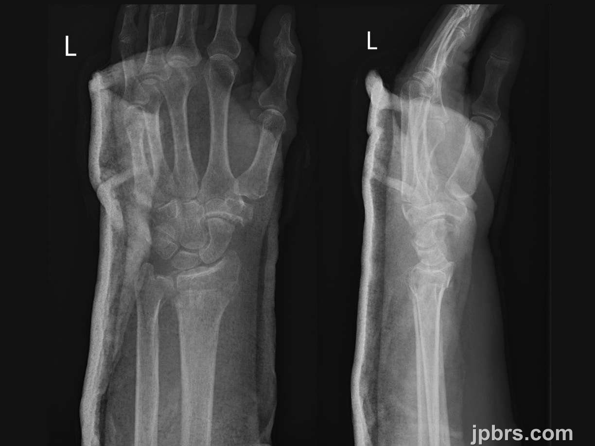
The radial inclination was measured on the AP view. It is defined as the angle between a line perpendicular to the long axis of the radial shaft (red line, (the blue line serves as a reference to guide placement of the red line)) and a line, representing the articular surface of the radius, drawn from the radial styloid to the ulnar edge of the radial articular surface (green line). This line should be placed at the central reference point of the articular surface (“the bottom of the cup”). The normal radial inclination is approximately 22 degrees. In this case, the radial inclination was 20 degrees.
2

Ulnar variance (blue line) refers to the difference in the relative lengths of the distal articular surfaces of the ulna (yellow line) and radius (orange line). There is considerable individual variability. The ulna is often 1 mm shorter than the radius. In this case, the ulna was 3 mm longer than the radius, which was considered unacceptable.
3

Before attempting closed reduction, a hematoma block was performed. It provides a highly effective form of analgesia prior to manipulation of the wrist.
The following materials were prepared: A drawing-up needle, injection needle, 20 ml syringe, lidocaine 1-2%, alcohol swabs, and optionally gauze. All needles were disposed in a sharps container after use.
4

The injection site should be slightly proximal to the fracture site. It may be easier to visualize the fracture site when comparing the clinical and radiological findings.
5

The injection site is disinfected twice.
6

The injection site should be placed slightly proximal to the fracture site. The needle is advanced until contact with the dorsal cortex is felt.
7

When the dorsal cortex is felt, the angulation is altered, so that the needle can slide along the cortex until the fracture line is reached.
8

Blood return on aspiration indicates that the needle tip is within the fracture line. The local anesthetic should be administered there. Inform the patient that they may feel pressure and pain. Take short breaks if necessary. Continue administration until slight resistance is felt, usually after 8-12 ml. If there is a fracture of the ulnar styloid, 2-3 ml can be injected subcutaneously at that site. Wait approximately 10 minutes before manipulating the fracture.
9

Chinese finger traps is a method that can be used alone (typically for impacted fractures and for patients with poor skin, who are at risk of wounds from manual reduction) or to induce muscle relaxation prior to manual reduction. There is no evidence that one method is superior to the other.
Ensure the hematoma block has taken effect. Select the appropriate size of fingertraps, attach them to first, second and third digits. Make sure the patient is positioned comfortably, and that there is sufficient space around the patient for manipulation and casting. The patient should lie in a supine position, with the upper arm perpendicular to the body, and the forearm perpendicular to the upper arm. The sandbag should be placed close to the elbow. Continue the traction for 20-30 minutes.
10

Manual reduction can be performed as a standalone procedure or following finger trap traction. Ensure the hematoma block has taken effect, and that sufficient personnel are available to assist.
Have someone provide countertraction by holding a towel wrapped around the upper arm, close to the elbow, which should be flexed at approximately 90 degrees. If there are not enough personnel, the towel can be secured to a fixed object. The clinician then grasps the patient’s hand, and at least one nurse should be ready to prepare the cast.
A useful tip is to hold on to first, second and third digits if the patient has fragile skin. But otherwise just get a firm grip.
11

Begin with traction to separate the fracture ends. Then pull the hand dorsally, as if attempting to make the dorsal angulation worse; this helps release impacted segments or entrapped structures.
12

Next, pull the hand volarly, to reestablish the physiological volar tilt, then maintain traction. Use the other hand to palpate the fracture site and detect a decrease in step-off.
13

If clinically satisfying, a dorsal below-elbow three-point support cast is applied while maintaining traction.
14

Once the cast has set sufficiently and the sides can no longer be manipulated, carefully and gradually release traction and place the arm in a sling until the cast is fully dry. A new radiograph should be obtained to ensure the position.
If the post-reduction position is acceptable, the patient continues with conservative treatment. However, due to manipulation of the wrist and the unstable fracture, a follow-up clinical assessment and radiograph should be performed after 10-12 days.
15
In this case, closed reduction was successful. Post-reduction radiograph showed an improved position, although not perfect. The angulation was 7 degrees dorsally, the length had improved, with the ulna now at -1. The inclination remained unchanged.
The position was regarded as acceptable. The patient was discharged and was prescribed paracetamol and ibuprofen for pain management.
The patient was referred for a follow-up in 7-10 days at which time the radiographic alignment will be reassessed. If the fracture remains in position, conservative treatment is continued; otherwise, operative treatment is indicated. In conservative management, the cast is maintained for five weeks. The patient should be introduced to edema prophylaxis and finger mobilization to prevent stiffness. Some patients may require physiotherapy after cast removal. The patient can expect improvement of wrist function over the first 6 months after cast removal; some patients may continue to improve function up to a year after removal.
Due to a low-energy trauma resulting in a wrist fracture, the patient should be referred for an osteoporosis workup.
How to cite this article
Maria Lerche Mortensen and Philip Månsson. Case 25 2026: Closed Reduction of a Distal Radius Fracture in the Emergency Department. Journal of Plastic, Breast & Reconstructive Surgery. 2026.
Open access. © 2026 The Author(s). Published by the Journal of Plastic, Breast & Reconstructive Surgery. Copyright to this case is shared between the author(s) and the journal; authors may reuse their own case for education, presentations and social media.
Keep reading
Related cases
Case 20 2026: Staged Excision and Delayed Primary Closure of a large Nodular Basal Cell Carcinoma of the Anterior Scalp
Mikkel Halborg Sørensen, Nanja Gotland Sundstrup, Christian Lyngsaa Lang
Keywords: Basal cell carcinoma, scalp reconstruction, staged excision, primary closure, scalp biomechanics. Authors: Mikkel Halborg Sørensen, MD; Nanja Gotland Sundstrup, MD; Christian Lyngsaa Lang, MD. Institution: Department of Plastic Surgery and Burns, Rigshospitalet, Copenhagen University Hos-pital, Denmark Abstract A 48-year-old woman was referred with a non-radically excised nodular basal cell carcinoma of the anterior scalp complicated by wound infection and dehiscence. Re-excision with 3 mm margins to the galea was performed under local anesthesia. Given local inflammation and uncertain margin status, delayed reconstruction was planned pending histopathological confirmation. After clear margins were verified, controlled subgaleal undermining allowed tension-reduced primary closure. This staged strategy ensured oncologic safety while preserving hair-bearing scalp and avoiding unnecessary flap reconstruction. Patient medical history A 48-year-old woman was referred after incomplete excision of a nodular basal cell carcinoma (nBCC) of the anterior scalp performed in private plastic surgery practice. Histology demonstrated tumor involvement of the lateral margins, while the deep margin was free of tumor. Postoperatively, wound rupture and infection developed. Microbiology showed sparse growth of Staphylococcus aureus, and antibiotic therapy was initiated. The patient was otherwise non-smoking and healthy, with well-treated asthma, as her only relevant comorbidity. Before and After Patient examination Clinical examination revealed a 22 × 19 mm anterior scalp defect with limited perifocal erythema and minimal purulent discharge. No regional lymphadenopathy was palpable. Pre-operative considerations Initial histopathology demonstrated nodular basal cell carcinoma with focal lateral margin involvement and a clear deep margin, without perineural invasion or other high-risk features. In accordance with current recommendations for low-risk BCC, re-excision with 3 mm c
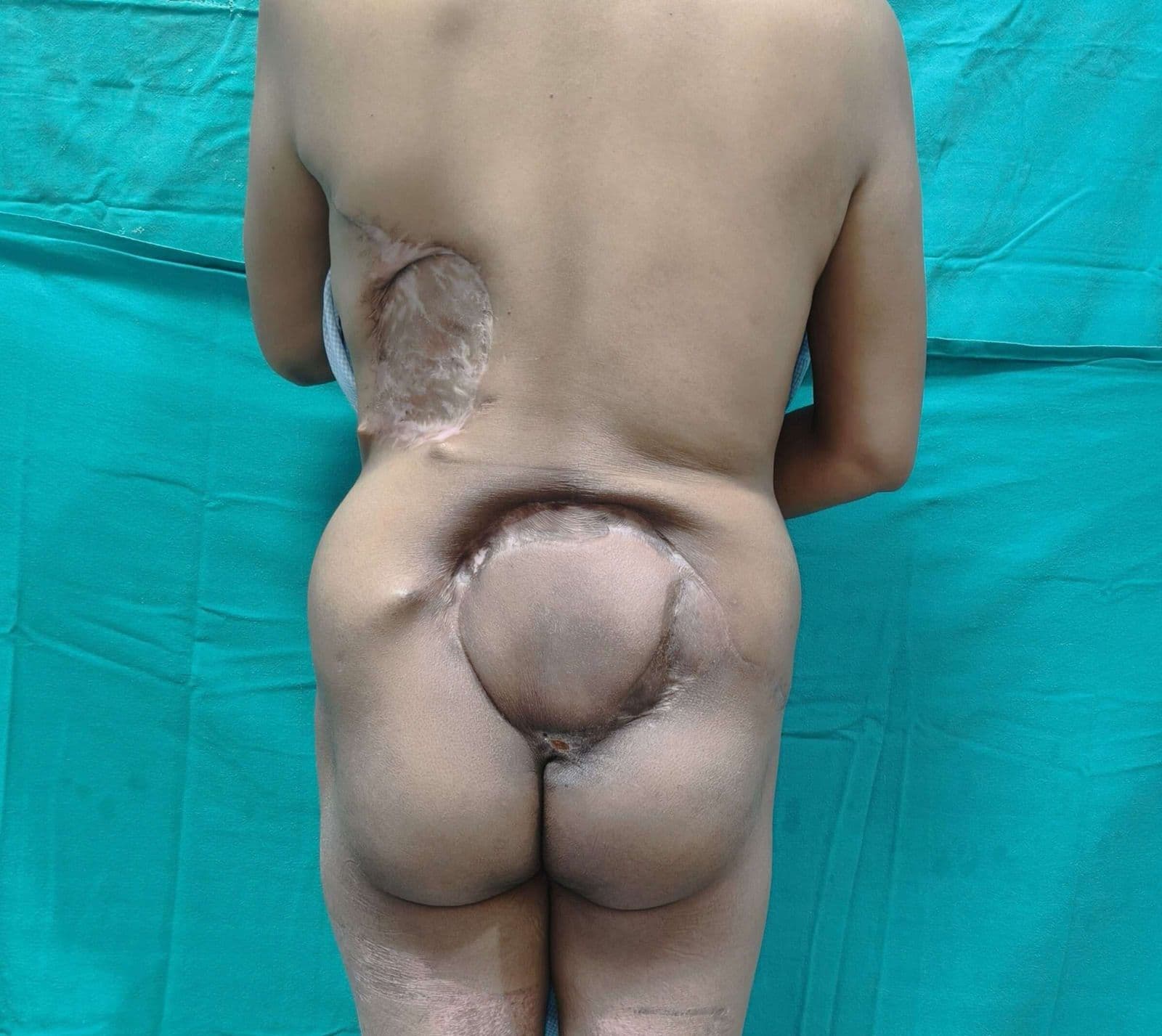
Case 22 2026: Superior Gluteal vessel: A rarely used recipient for free flap reconstruction of lumbosacral defects: a case report.
Tushar Dutta, Arun PS, Swati Sattavan
Keywords: Superior gluteal vessel, free flap reconstruction, lumbosacral defects Authors: 1. Tushar Dutta, Consultant Plastic Surgeon; 2. Arun PS, Consultant Surgical Oncologist; 3. Swati Sattavan, Consultant Surgical Oncologist; Institute: State Cancer Institute, Guwahati, Assam, India Abstract Lumbosacral defects that are too large for local flap reconstruction should be considered for microvascular free tissue transfer. However, lack of suitable recipient vessels in the area makes the task technically difficult. Options such as thoracodorsal and inferior gluteal vessels extended with vein grafts and AV loops have been described. Each option has its own drawbacks. Despite having a short course at its origin, the superior gluteal vessel is a viable alternative. We describe here a case where the vessel was dissected out with adequate length and used as a recipient for free flap reconstruction. Patient medical history The patient was a 41-year-old lady with no known comorbidities. She presented to us with a large lumbosacral mass that had been growing for one year. Before visiting our center, she had been operated twice at other institutions and had recurrences after each occasion. During the last three months the mass had started to grow rapidly and caused ulceration. There was no pain or fever. There was no neurological involvement of lower limbs. Patient had an ECOG status 1. Before and After Patient examination A 17x 22 cm mass was noted encompassing the left part of lumbosacral region and crossing the midline to the opposite side. The superior limit of the lesion was at the L3 vertebra and inferior extent was near the coccyx. Skin ulceration was noted at the center of the mass. On palpation, the mass was warm to touch, hard in consistency and immobile. Skin was fixed and fixity was also noted to the underlying muscle. No tenderness was elicited. Surrounding possible donor sites for flap harvest showed no scars. Pre-operative considerations Preoperative biopsy co
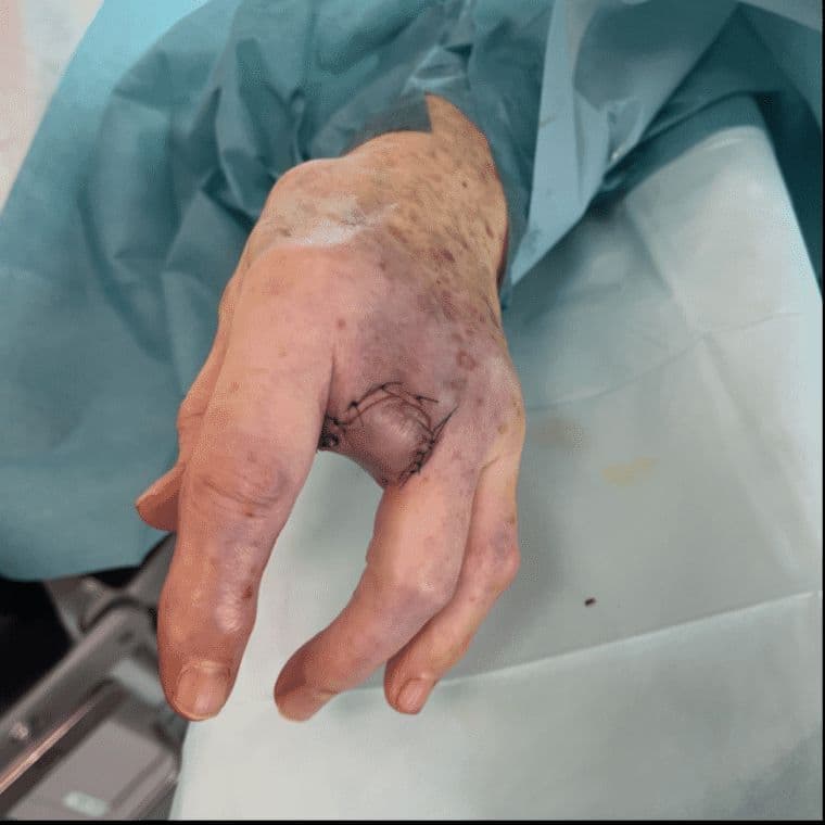
Case 23 2026: “Minimally Invasive Finger Amputation Using a Volar ‘Toilet seat’ Flap for Invasive Squamous Cell Carcinoma”
Claes Hannibal Killerich, Nikolaj Warming
Keywords: Squamous cell carcinoma; finger amputation; volar flap; local reconstruction; traction neurectomy; hand surgery. Authors: Claes Hannibal Killerich, Nikolaj Warming Abstract A 93-year-old woman presented with a chronic non-healing dorsal finger wound initially diagnosed as actinic keratosis. Repeat biopsy revealed invasive squamous cell carcinoma with joint and bone involvement. Due to the extent of invasion, distal amputation of the third finger was required. Reconstruction was performed using a volar “toilet seat” flap under digital nerve block to minimize surgical burden. The procedure achieved clear margins, preserved stump length, and avoided donor-site morbidity. This case highlights the importance of early re-biopsy of non-healing lesions and adapting surgical strategy to patient age, function, and comorbidity. Patient medical history A 93-year-old woman presented with a wound on the dorsal aspect of the proximal phalanx of the left third finger. A primary biopsy revealed actinic keratosis, and the lesion was treated several times with curettage and electrodesiccation by a private dermatologist. Despite repeated treatments under professional supervision, the wound failed to heal.Due to the persistent non-healing nature of the lesion, a repeat biopsy was performed, confirming the diagnosis of squamous cell carcinoma (SCC). The patient was referred to the Department of Plastic Surgery, Aalborg University Hospital, where a primary excision was performed with a 7 mm surgical margin. Histopathological examination demonstrated invasive tumor growth involving both joint and bone. The defect was subsequently reconstructed using a full-thickness skin graft. Before and After Patient examination The left third finger showed a healed full-thickness skin graft on the dorsal aspect of the proximal phalanx, covering the proximal interphalangeal (PIP) joint. There was no visible residual tumor, and no palpable lymphadenopathy in the left cubital fossa or axillary re