Case 20 2026: Staged Excision and Delayed Primary Closure of a large Nodular Basal Cell Carcinoma of the Anterior Scalp
Mikkel Halborg Sørensen, Nanja Gotland Sundstrup, Christian Lyngsaa Lang
Published · May 28, 2026
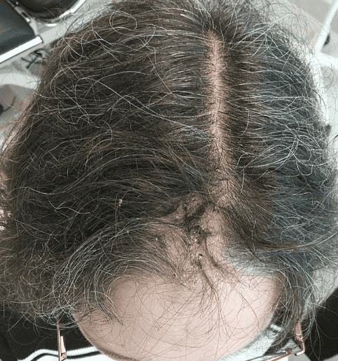
Keywords: Basal cell carcinoma, scalp reconstruction, staged excision, primary closure, scalp biomechanics. Authors: Mikkel Halborg Sørensen, MD; Nanja Gotland Sundstrup, MD; Christian Lyngsaa Lang, MD. Institution: Department of Plastic Surgery and Burns, Rigshospitalet, Copenhagen University Hos-pital, Denmark Abstract A 48-year-old woman was referred with a non-radically excised nodular basal cell carcinoma of the anterior scalp complicated by wound infection and dehiscence. Re-excision with 3 mm margins to the galea was performed under local anesthesia. Given local inflammation and uncertain margin status, delayed reconstruction was planned pending histopathological confirmation. After clear margins were verified, controlled subgaleal undermining allowed tension-reduced primary closure. This staged strategy ensured oncologic safety while preserving hair-bearing scalp and avoiding unnecessary flap reconstruction. Patient medical history A 48-year-old woman was referred after incomplete excision of a nodular basal cell carcinoma (nBCC) of the anterior scalp performed in private plastic surgery practice. Histology demonstrated tumor involvement of the lateral margins, while the deep margin was free of tumor. Postoperatively, wound rupture and infection developed. Microbiology showed sparse growth of Staphylococcus aureus, and antibiotic therapy was initiated. The patient was otherwise non-smoking and healthy, with well-treated asthma, as her only relevant comorbidity. Before and After Patient examination Clinical examination revealed a 22 × 19 mm anterior scalp defect with limited perifocal erythema and minimal purulent discharge. No regional lymphadenopathy was palpable. Pre-operative considerations Initial histopathology demonstrated nodular basal cell carcinoma with focal lateral margin involvement and a clear deep margin, without perineural invasion or other high-risk features. In accordance with current recommendations for low-risk BCC, re-excision with 3 mm c
References
- Gulleth Y, Goldberg N, Silverman RP, Gastman BR. What is the best surgical margin for a Basal cell carcinoma: a meta-analysis of the literature PRS. 2010 oct.; 126 (4):1222-31
- Leedy JE, Janis JE, Rohrich RJ. Reconstruction of acquired scalp defects: an algorithmic approach. Plastic and Reconstructive Surgery. 2005;116(4):54e–72e. DOI: 10.1097/01.prs.0000179188.75076.9b
- Beasley NJ, Gilbert RW. Scalp and forehead reconstruction. Clinics in Plastic Surgery. 2005;32(2):203–215. DOI: 10.1016/j.cps.2004.11.002