Case 2 - 2025: Reconstruction of an extensive foot defect using a combined sensory split latissimus dorsi and serratus anterior muscle flap
Dr. med. Luisa Lotter, PD
Published · April 2, 2025
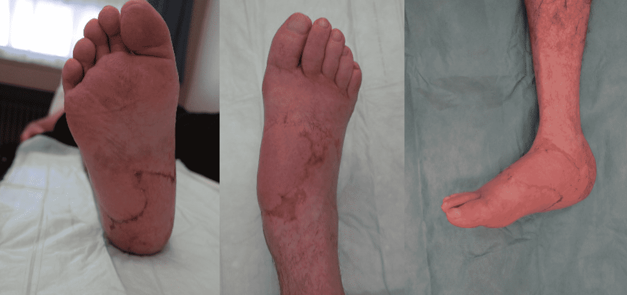
Keywords: Lower extremity; Flap; Reconstruction; Soft tissue; Trauma; Microsurgery; Foot; Latissimus Dorsi ; Authors: Dr. med. Luisa Lotter, Dr. med. Ilja Käch, PD Dr. med. Volker Schmidt. Institution: Kantonsspital St. Gallen, Switzerland Abstract A 67-year-old male patient sustained a complex foot injury with an open Lisfranc dislocation of the medial cuneiform bone and shaft fractures of the 2nd to 4th metatarsals in July 2024, as a result of a crush trauma. After initial osteosynthetic treatment, a pronounced necrosis developed in the area of the hindfoot and forefoot over time and an implant-associated wound infection. The patient was then transferred to our tertiary care hospital for specialized defect reconstruction. At first radical necrosectomy was performed, resulting in a 10 x 12 cm soft-tissue defect in the heel area, which extended medially and laterally. In the area of the forefoot, there was also a full-thickness wound measuring 4 x 3 cm with exposed osteosynthetic material, bone, and tendon. Patient medical history The patient has no pre-existing conditions. Blood tests on admission showed a blood glucose level and HbA1c value within the normal range. An angiography was performed, revealing a regular three-vessel supply to the affected lower right leg. Before and After Patient examination Upon admission to our hospital, on assessment, the 67-year-old male patient was in good general health with a slim nutritional status. On the heel area of the right foot was a 10 x 12 cm soft-tissue defect, which extended medially and laterally. In the area of the forefoot, there was also a full-thickness wound measuring 4 x 3 cm with exposed osteosynthetic material, bone, and tendon. Blood tests on admission showed a blood glucose level and HbA1c value within the normal range. An angiography was performed, revealing a regular three-vessel supply to the affected lower right leg. Pre-operative considerations The reconstruction of complex soft tissue injuries with exp
Step-by-step
- Step 1
- Step 2
- Step 3
- Step 4
References
- Harvey Chim, Rachel Cohen-Shohet, Mariel M McLaughlin, Tosan Ehanire. Function-Sparing Free Split Latissimus Dorsi Flap for Lower-Extremity Reconstruction: Five-Year Consecutive Single-Surgeon Series. J Bone Joint Surg Am . 2020 Oct 7;102(19):1714-1723. doi: 10.2106/JBJS.20.00022
- Thomas Albert, Stéphane Guero, Clément Deranque, Pascal Rousseau. Combined free flap of muscle-sparing latissimus dorsi and serratus anterior in the repair of child’s traumatic foot injury: a case report. Pan Afr Med J. 2024 Jul 9;48:91. doi: 10.11604/pamj.2024.48.91.43952