Case 19 2026: Split Perforator Flaps for bilateral ankle defects: A modern microsurgical approach to preserve function and reduce secondary thinning operations
Luisa Lotter¹, Marta Jezierska¹, Ilja W. Käch¹, Volker J. Schmidt¹
Published · May 28, 2026
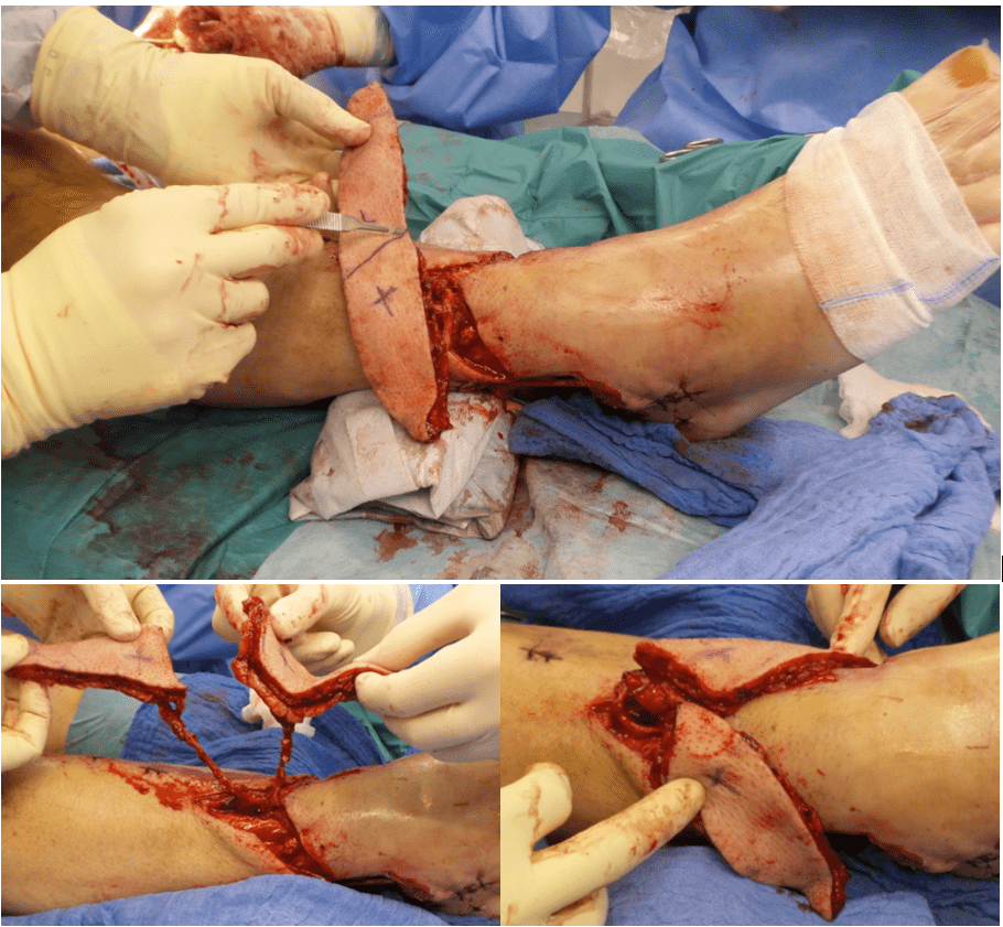
Keywords: Lower extremity; Orthoplastic Reconstruction; Split flap; microsurgery Authors: Luisa Lotter¹, Marta Jezierska¹, Ilja W. Käch¹, Volker J. Schmidt¹; ¹Department of Plastic Surgery and Handsurgery, HOCH, Health Ostschweiz, Cantonal Hospital St.Gallen, St.Gallen, Switzerland Abstract 46-year-old man presented with two full-thickness soft-tissue defects of the distal right lower leg (medial 7 × 4 cm, lateral 10 × 5 cm) with exposed tendons, osteosynthesis material, and bone after a Gustilo grade IIIb open tibial and fibular fracture. During initial orthopedic stabilization, interdisciplinary orthoplastic assessment and planning were performed. Because of exposed functional structures and two spatially separated defects with healthy anterior skin between them, microsurgical free-tissue reconstruction was chosen to provide stable coverage while preserving the anterior skin and preventing flap-related functional impairment. Reconstruction was achieved using a split anterolateral thigh (ALT) flap based on two independent perforators, enabling simultaneous coverage of both defects with a single vascular pedicle. Patient medical history The patient’s medical history was significant for type 2 diabetes mellitus, two-vessel coronary artery disease, and obesity. Before and After Patient examination Clinical assessment revealed two full-thickness defects at the distal lower leg: • medial defect: 7 × 4 cm with exposed tendons • lateral defect: 10 × 5 cm with exposed bone and osteosynthesis material Both wounds showed non-viable soft-tissue components requiring radical surgical debridement prior to reconstruction. An CT-angiography was performed, revealing a regular three-vessel supply to the affected lower right leg. Preoperative color-duplex-ultrasound was routinely performed to identify the ALT perforator pattern and to verify, if a two-perforator split ALT-fashion is possible. Due to our tertiary orthoplastic program final interdisciplinary reconstruction was schedule
References
- Donor site in anterolateral thigh (ALT) free flaps: A systematic review of closure techniques and introduction of a management algorithm Chang, Chad et al. Journal of Plastic, Reconstructive & Aesthetic Surgery, Volume 105, 243 – 259
- Split anterolateral thigh free flaps for bilateral open fracture ankle defects: A one donor, two flap solution, Wilson, Elizabeth et al. JPRAS Open, Volume 46, 2025, Pages 61-64