
Case 12 2026: Closure of defect on ala nasi with modified shark island flap
Mickie Bang Christensen (MD)1, Magnus Avnstorp (MD)1
- 1Department of Plastic- & Breast Surgery, Zealand University Hospital Roskilde, Denmark
Abstract
Keywords: Shark island flap, facial skin defect, BCC, nasal skin cancer, nasal reconstruction, reconstructive surgery Authors: Mickie Bang Christensen (MD), Magnus Avnstorp (MD), Department of Plastic- & Breast Surgery, Zealand University Hospital Roskilde, Denmark Abstract This case presents the use of a modified shark island flap for reconstruction of a defect on the ala nasi following excision of a basal cell carcinoma in a 85-year old male. Due to the tumor size and location, direct closure was not feasible. The shark island flap was chosen as the reconstructive approach. Surgery was done under local anesthesia. Excision of the tumor was done with a 3 mm margin. Histology from frozen sections showed clear margins without malignancy. Postoperative follow up at both one week and two months demonstrated a satisfactory cosmetic outcome and no signs of recurrence. Patient medical history 85-year old male with a medical history of atoxic goiter, hypertension, Parkinson's disease, melanoma and multiple skin carcinomas, was referred with 3 facial skin carcinomas (dorsum nasi, right ala nasi and left side of the upper lip). This case focuses on the carcinoma on the ala nasi. The biopsy showed basal cell carcinoma of nodular subtype. The patient had years prior been operated with a bilobed flap due to a skin carcinoma on the right dorsum nasi, which had resulted in bulging of the right side of the dorsum. Before and After Patient examination Well preserved male with poor skin quality. On clinical examination, a tumor measuring 6 x 5 mm and 2 mm elevation from the surface of the skin was found on the right ala nasi, involving the nasal sulcus. Clinically not adherent to underlying tissue. There were no palpable regional lymph nodes. Pre-operative considerations Surgery and radiation were discussed with the patient. The patient preferred surgery. Due to the tumor size and location, direct closure was not feasible. FTSG would delete the alar-facial sulcus, create a big,
 BeforeAfter
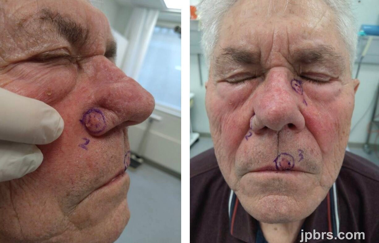
85-year old male with a medical history of atoxic goiter, hypertension, Parkinson's disease, melanoma and multiple skin carcinomas, was referred with 3 facial skin carcinomas (dorsum nasi, right ala nasi and left side of the upper lip). This case focuses on the carcinoma on the ala nasi. The biopsy showed basal cell carcinoma of nodular subtype. The patient had years prior been operated with a bilobed flap due to a skin carcinoma on the right dorsum nasi, which had resulted in bulging of the right side of the dorsum.

Excision with frozen sections and a 3 mm safety margin resulted in a defect measuring 9 x 8 mm. The tumour was marked by needle at 12’o clock, superiorly. Frozen sections showed clear margins.
The shark island flap is a myocutaneus flap first described by Cvancara and Wentzell in 2006 and obtained the name due to its shark-like shape. It is indicated for reconstruction of the alar and perialar defects, since it involves a rotation that gives rise to the formation of an inverted cone that recreates the alar sulcus. The flap derives its blood supply from the levator labii superioris muscle and requires careful dissection to preserve the vascular pedicle.
I. The flap is drawn by measuring the width of the defect from the lateral alar sulcus to the medial edge of the defect. A matching width of a short limb of the flap is measured. The long limp of the flap goes down the cheek with the inferior portion of the island pedicle. Then the medial edge of the flap is drawn on the nasolabial fold.
II. Then the subcutaneous pedicled flap is incised. Carefully undermining is done in order to preserve musculature at the base of the pedicle on the short limb.
III. The short limb is rotated 90 degrees, perpendicular to the melolabial part of the flap, thereby recreating the alar sulcus. The first deep suture pulls the upper lateral part towards the nasofacial sulcus, allowing the short limb to drape down into the defect. The second deep suture pulls the short limb down to the inferior border of the defect..
IV. The remainder of the flap is carefully sutured deeply and superficially, avoiding too many deep sutures to minimize the risk of necrosis. (1)

The flap was drawn on the patient as shown on the drawing from step 1.
2

Subcutaneous dissection is carefully done in order not to damage the facial artery and its branches. The flap receives its nervous innervation from n. infraorbitalis and rami buccales. Adequate pedicle size is important in order to avoid necrosis. Furthermore, it’s crucial to identify the alar crease and the nasolabial fold and draw the flap according to those anatomical landmarks.
3

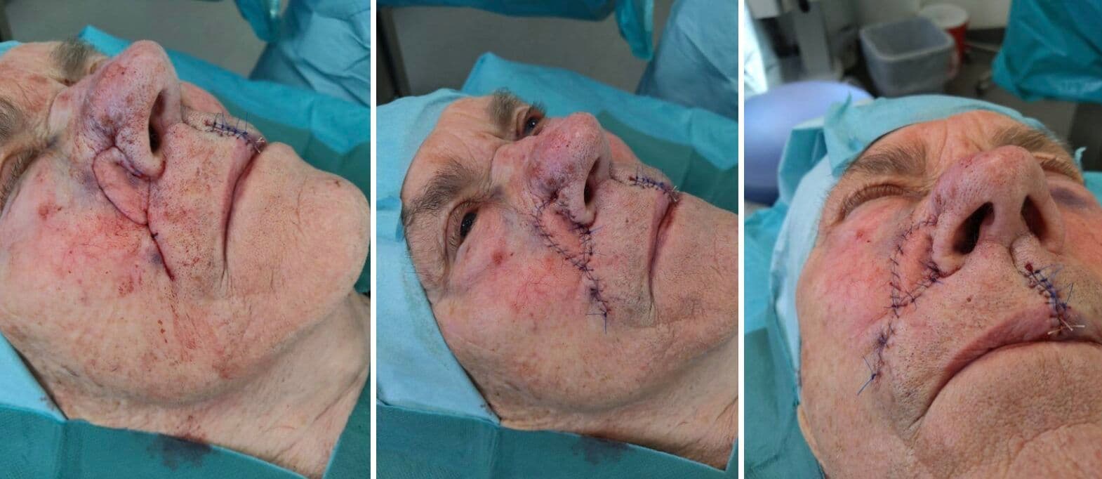
The flap was carefully incised and subcutaneously dissected in order to preserve the musculature at the base of the pedicle.
4
The short limb of the flap is lifted towards the nose, folded and then sutured with 5-0 absorbable polyfilament sutures. The inferior part of the flap-defect is closed, also with 5-0 sutures, successfully re-creating the nasolabial fold. The long limb of the flap is trimmed/shortened, making it more well-fitted for the defect.
5

The remaining part is sutured with a few 5-0 absorbable polyfilament sutures. Finally, the skin is sutured with 5-0 continuous non-absorbable monofilament sutures, ensuring precise adaptation of the skin edges.
6
The patient presented with discrete hematoma on the cheek. The flap, fully healed, with a small amount of central bulging. Normal capillary response. The patient had no complaints
7

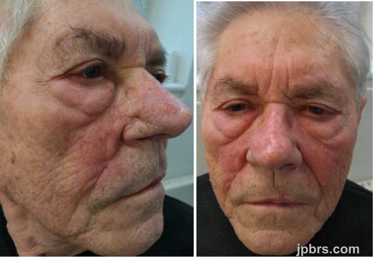
The flap was seen fully healed and the central bulging gone. The alar crease and the nasolabial fold had been recreated due to the flap design, blending well with the contralateral side. No signs of recurrence. The patient was satisfied with the cosmetic outcome.
8

9
The patient was advised to keep the head elevated and refrain from consuming hot beverages and food 1-2 days after surgery. Additionally, no strenuous physical activities or swimming until removal of the sutures 7 days post-operatively.
How to cite this article
Mickie Bang Christensen (MD), Magnus Avnstorp (MD). Case 12 2026: Closure of defect on ala nasi with modified shark island flap. Journal of Plastic, Breast & Reconstructive Surgery. 2026.
Open access. © 2026 The Author(s). Published by the Journal of Plastic, Breast & Reconstructive Surgery. Copyright to this case is shared between the author(s) and the journal; authors may reuse their own case for education, presentations and social media.
Keep reading
Related cases
Case 20 2026: Staged Excision and Delayed Primary Closure of a large Nodular Basal Cell Carcinoma of the Anterior Scalp
Mikkel Halborg Sørensen, Nanja Gotland Sundstrup, Christian Lyngsaa Lang
Keywords: Basal cell carcinoma, scalp reconstruction, staged excision, primary closure, scalp biomechanics. Authors: Mikkel Halborg Sørensen, MD; Nanja Gotland Sundstrup, MD; Christian Lyngsaa Lang, MD. Institution: Department of Plastic Surgery and Burns, Rigshospitalet, Copenhagen University Hos-pital, Denmark Abstract A 48-year-old woman was referred with a non-radically excised nodular basal cell carcinoma of the anterior scalp complicated by wound infection and dehiscence. Re-excision with 3 mm margins to the galea was performed under local anesthesia. Given local inflammation and uncertain margin status, delayed reconstruction was planned pending histopathological confirmation. After clear margins were verified, controlled subgaleal undermining allowed tension-reduced primary closure. This staged strategy ensured oncologic safety while preserving hair-bearing scalp and avoiding unnecessary flap reconstruction. Patient medical history A 48-year-old woman was referred after incomplete excision of a nodular basal cell carcinoma (nBCC) of the anterior scalp performed in private plastic surgery practice. Histology demonstrated tumor involvement of the lateral margins, while the deep margin was free of tumor. Postoperatively, wound rupture and infection developed. Microbiology showed sparse growth of Staphylococcus aureus, and antibiotic therapy was initiated. The patient was otherwise non-smoking and healthy, with well-treated asthma, as her only relevant comorbidity. Before and After Patient examination Clinical examination revealed a 22 × 19 mm anterior scalp defect with limited perifocal erythema and minimal purulent discharge. No regional lymphadenopathy was palpable. Pre-operative considerations Initial histopathology demonstrated nodular basal cell carcinoma with focal lateral margin involvement and a clear deep margin, without perineural invasion or other high-risk features. In accordance with current recommendations for low-risk BCC, re-excision with 3 mm c
Case 22 2026: Superior Gluteal vessel: A rarely used recipient for free flap reconstruction of lumbosacral defects: a case report.
Tushar Dutta, Arun PS, Swati Sattavan
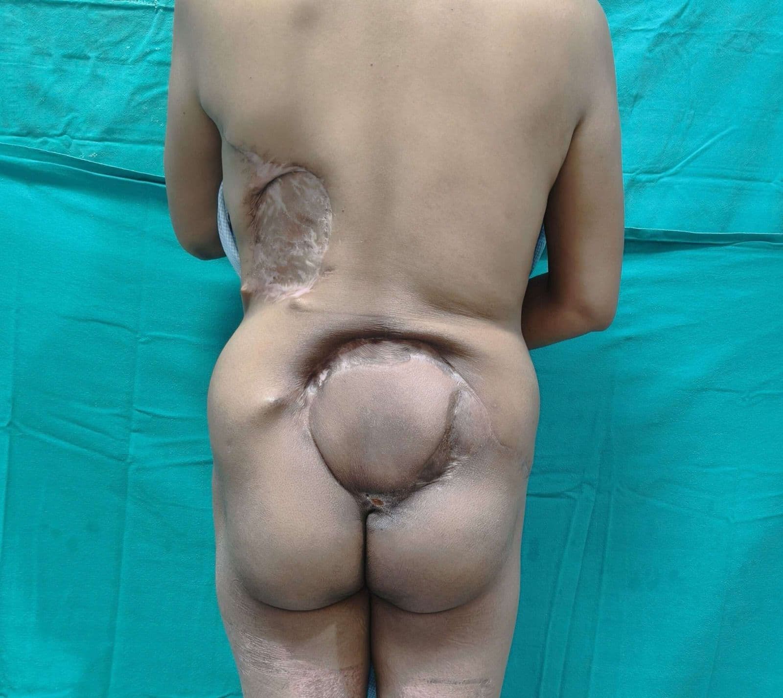
Keywords: Superior gluteal vessel, free flap reconstruction, lumbosacral defects Authors: 1. Tushar Dutta, Consultant Plastic Surgeon; 2. Arun PS, Consultant Surgical Oncologist; 3. Swati Sattavan, Consultant Surgical Oncologist; Institute: State Cancer Institute, Guwahati, Assam, India Abstract Lumbosacral defects that are too large for local flap reconstruction should be considered for microvascular free tissue transfer. However, lack of suitable recipient vessels in the area makes the task technically difficult. Options such as thoracodorsal and inferior gluteal vessels extended with vein grafts and AV loops have been described. Each option has its own drawbacks. Despite having a short course at its origin, the superior gluteal vessel is a viable alternative. We describe here a case where the vessel was dissected out with adequate length and used as a recipient for free flap reconstruction. Patient medical history The patient was a 41-year-old lady with no known comorbidities. She presented to us with a large lumbosacral mass that had been growing for one year. Before visiting our center, she had been operated twice at other institutions and had recurrences after each occasion. During the last three months the mass had started to grow rapidly and caused ulceration. There was no pain or fever. There was no neurological involvement of lower limbs. Patient had an ECOG status 1. Before and After Patient examination A 17x 22 cm mass was noted encompassing the left part of lumbosacral region and crossing the midline to the opposite side. The superior limit of the lesion was at the L3 vertebra and inferior extent was near the coccyx. Skin ulceration was noted at the center of the mass. On palpation, the mass was warm to touch, hard in consistency and immobile. Skin was fixed and fixity was also noted to the underlying muscle. No tenderness was elicited. Surrounding possible donor sites for flap harvest showed no scars. Pre-operative considerations Preoperative biopsy co
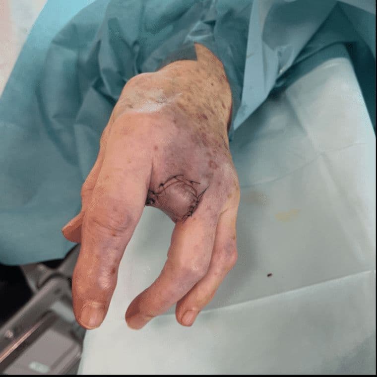
Case 23 2026: “Minimally Invasive Finger Amputation Using a Volar ‘Toilet seat’ Flap for Invasive Squamous Cell Carcinoma”
Claes Hannibal Killerich, Nikolaj Warming
Keywords: Squamous cell carcinoma; finger amputation; volar flap; local reconstruction; traction neurectomy; hand surgery. Authors: Claes Hannibal Killerich, Nikolaj Warming Abstract A 93-year-old woman presented with a chronic non-healing dorsal finger wound initially diagnosed as actinic keratosis. Repeat biopsy revealed invasive squamous cell carcinoma with joint and bone involvement. Due to the extent of invasion, distal amputation of the third finger was required. Reconstruction was performed using a volar “toilet seat” flap under digital nerve block to minimize surgical burden. The procedure achieved clear margins, preserved stump length, and avoided donor-site morbidity. This case highlights the importance of early re-biopsy of non-healing lesions and adapting surgical strategy to patient age, function, and comorbidity. Patient medical history A 93-year-old woman presented with a wound on the dorsal aspect of the proximal phalanx of the left third finger. A primary biopsy revealed actinic keratosis, and the lesion was treated several times with curettage and electrodesiccation by a private dermatologist. Despite repeated treatments under professional supervision, the wound failed to heal.Due to the persistent non-healing nature of the lesion, a repeat biopsy was performed, confirming the diagnosis of squamous cell carcinoma (SCC). The patient was referred to the Department of Plastic Surgery, Aalborg University Hospital, where a primary excision was performed with a 7 mm surgical margin. Histopathological examination demonstrated invasive tumor growth involving both joint and bone. The defect was subsequently reconstructed using a full-thickness skin graft. Before and After Patient examination The left third finger showed a healed full-thickness skin graft on the dorsal aspect of the proximal phalanx, covering the proximal interphalangeal (PIP) joint. There was no visible residual tumor, and no palpable lymphadenopathy in the left cubital fossa or axillary re