
Case 16 2026: Severe Foot Degloving Injury Following a Forklift Crush: A Limb-Salvage Case
Cecilie Jansen1, Emil Villiam Holm-Rasmussen1, Mette Stueland Wolthers1
- 1University Hospital, Rigshospitalet, Denmark
Abstract
Keywords: Degloving injury, (decollement), foot trauma, limb salvage, fluorescence angiography, negative pressure wound therapy. Authors: Cecilie Jansen, Emil Villiam Holm-Rasmussen, Mette Stueland Wolthers. Copenhagen University Hospital, Rigshospitalet, Denmark. Abstract A 30-year-old previously healthy man was admitted to The Trauma Centre at Copenhagen University Hospital, Rigshospitalet after his left foot was crushed by a 1.5-ton forklift at work. He sustained a large open fracture and an extensive circumferential degloving injury involving the foot and ankle, while distal perfusion remained intact. Computed Tomography (CT) demonstrated multiple intra-articular fractures with suspected Lisfranc injury and no vascular injury on CT angiography. Following orthopedic stabilization, plastic surgical management focused on debridement, tissue viability assessment, skin and heel-pad preservation, and limb salvage. This case highlights the challenges of severe foot degloving and multidisciplinary limb-salvage management. Patient medical history The patient was previously healthy, had no significant past medical history, and was a non-smoker. Before and After Patient examination On arrival at The Trauma Centre at Copenhagen University Hospital, Rigshospitalet, the patient was hemodynamically stable but slightly pale. He reported paresthesia in the left foot, but gross sensation was preserved. Clinical examination revealed an approximately 15 cm open wound laterally on the left foot with exposed bone and tendon. A palpable pulse was present in the dorsalis pedis artery, whereas the posterior tibial artery pulse was not palpable. Capillary refill and sensation were preserved. A trauma CT scan with intravenous contrast, including CT angiography of the lower leg was performed. Imaging demonstrated multiple intra-articular fractures distal to the talus with widening between the second metatarsal and medial cuneiform, raising suspicion of a Lisfranc injury. Air was present in
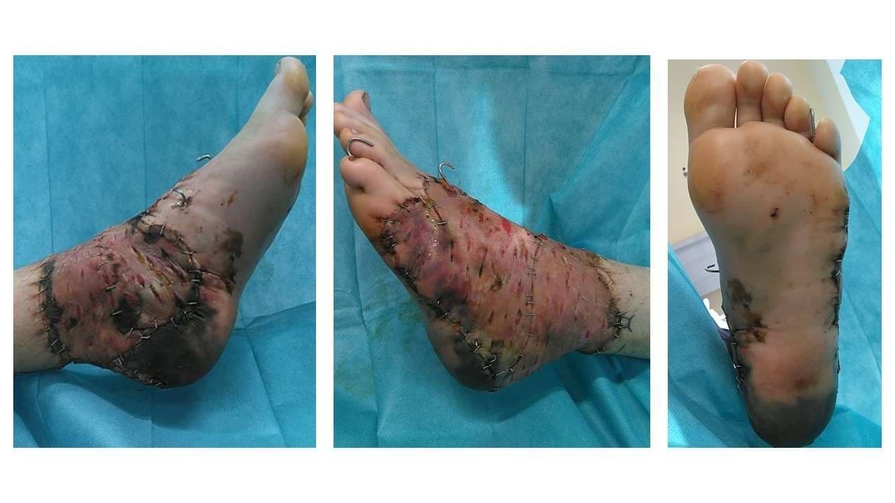
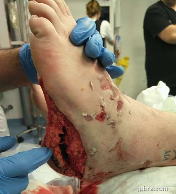
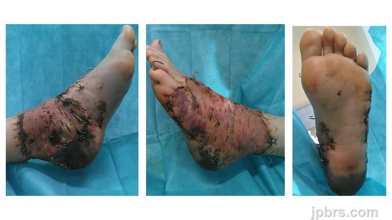 BeforeAfter
The patient was previously healthy, had no significant past medical history, and was a non-smoker.

Intraoperative examination revealed a large open degloving injury, predominantly circumferential around the ankle, extending approximately one handbreadth proximally. The injury continued distally over the dorsum of the foot and around both malleoli. The plantar surface showed partial avulsion involving the heel pad and the medial and lateral borders of the foot. Communication between the open wound and the degloving cavity was noted at the posteromedial heel and ankle region. Areas of crush injury and skin discoloration were present, particularly on the dorsum and medial aspect of the heel, indicating a risk of secondary necrosis. Palpable dorsalis pedis artery pulse was present, whereas the posterior tibial artery pulse was uncertain.

Intraoperative fluorescence angiography with intravenous administration of Indocyanine green
(ICG) dye demonstrated reduced or absent perfusion in a circumferential zone around the ancle, approximately 10 cm wide, extending from lateral to medial malleolus and distally over the dorsum of the foot. Perfusion was relatively preserved on the plantar aspect, including the heel pad. Clearly non-viable and undermined skin was marked and excised, leaving a 1 cm border, which was harvested as full-thickness. Systematic revision ensured complete removal of devitalized tissue while preserving viable structures.
2

Hemostasis was obtained. The plantar surface and heel pad were repositioned and anchored to the deep fascia and plantar structures using 2-0 Vicryl to reduce dead space and optimize the chance for tissue survival.
3

The avulsed tissue was harvested, trimmed/defatted, and meshed to a large full-thickness skin graft.
4

The full-thickness skin graft was applied over the foot and ancle and secured with staples. A 14-F suction drain was placed.
5

The graft was dressed with Cuticerin, and surrounding skin was prepared with adhesive film. A black sponge was applied over the grafted area and connected to continuous negative pressure at 125 mmHg.
6

Five days postoperatively, the full-thickness skin graft, particularly in the caudal/posterior region, showed mild maceration with some open mesh interstices. Overall graft take is difficult to assess at this stage. Most of the plantar surface appears pink with good capillary refill and preserved sensation. The heel pad and surrounding area remain somewhat questionable; the skin appears slightly pale to bluish-purple but is warm to touch. Toe mobility is intact, with preserved capillary refill and sensation throughout.
The full-thickness skin graft was then re-covered with a Peel and Place Dressing and NPWT was continued. A transparent Tegaderm dressing was applied to the heel to allow monitoring of the underlying skin while providing gentle compression to help reapproximate the plantar skin and heel pad to the underlying tissue.
7

Ten days postoperatively, the full-thickness skin graft, particularly in the caudal/posterior region, showed signs of mild maceration. Overall graft take was estimated at approximately 90%. Most of the plantar surface still appears pink with good capillary refill and preserved sensation. The heel pad and surrounding area appears pink and warm.
The full-thickness skin graft and heel pad was then covered with nitrofurantoin impregnated gauze, Jelonet dressing and a compression bandage with daily dressing changes and air dry to prevent the development of maceration.
8

Fourteen days postoperatively a line of demarcation is slowly beginning to appear at the heel pad. There are early signs of marginal necrosis at the site of the full-thickness skin graft. Overall graft take was estimated at approximately 90%. Most of the plantar surface still appears pink with good capillary refill and preserved sensation. The demarcation will be monitored for the next months, with a conservative approach.
9
The post-operative plan included NPWT dressing changes every 5–7 days. The left foot and lower limb were kept elevated at all times, with pressure avoided over the heel, and were kept warm using padding and gauze with additional covering as needed. The patient remained on strict bed rest for the first three days, followed by wheelchair mobilization with the leg elevated until the first dressing change. Weight-bearing on the affected foot was restricted for six weeks according to orthopedic guidelines. Continuous suction drainage was maintained until NPWT removal.
Once the skin graft is considered well healed, or near complete healed, the patient can be discharged home with daily dressing changes at home and a follow-up every 14th day at the department of plastic surgery, to assess healing, function, and any need for secondary revision. The expectation is that the heel will progress toward spontaneous healing after demarcation.
How to cite this article
Cecilie Jansen, Emil Villiam Holm-Rasmussen, Mette Stueland Wolthers. Case 16 2026: Severe Foot Degloving Injury Following a Forklift Crush: A Limb-Salvage Case. Journal of Plastic, Breast & Reconstructive Surgery. 2026.
Open access. © 2026 The Author(s). Published by the Journal of Plastic, Breast & Reconstructive Surgery. Copyright to this case is shared between the author(s) and the journal; authors may reuse their own case for education, presentations and social media.
Keep reading
Related cases
Case 20 2026: Staged Excision and Delayed Primary Closure of a large Nodular Basal Cell Carcinoma of the Anterior Scalp
Mikkel Halborg Sørensen, Nanja Gotland Sundstrup, Christian Lyngsaa Lang
Keywords: Basal cell carcinoma, scalp reconstruction, staged excision, primary closure, scalp biomechanics. Authors: Mikkel Halborg Sørensen, MD; Nanja Gotland Sundstrup, MD; Christian Lyngsaa Lang, MD. Institution: Department of Plastic Surgery and Burns, Rigshospitalet, Copenhagen University Hos-pital, Denmark Abstract A 48-year-old woman was referred with a non-radically excised nodular basal cell carcinoma of the anterior scalp complicated by wound infection and dehiscence. Re-excision with 3 mm margins to the galea was performed under local anesthesia. Given local inflammation and uncertain margin status, delayed reconstruction was planned pending histopathological confirmation. After clear margins were verified, controlled subgaleal undermining allowed tension-reduced primary closure. This staged strategy ensured oncologic safety while preserving hair-bearing scalp and avoiding unnecessary flap reconstruction. Patient medical history A 48-year-old woman was referred after incomplete excision of a nodular basal cell carcinoma (nBCC) of the anterior scalp performed in private plastic surgery practice. Histology demonstrated tumor involvement of the lateral margins, while the deep margin was free of tumor. Postoperatively, wound rupture and infection developed. Microbiology showed sparse growth of Staphylococcus aureus, and antibiotic therapy was initiated. The patient was otherwise non-smoking and healthy, with well-treated asthma, as her only relevant comorbidity. Before and After Patient examination Clinical examination revealed a 22 × 19 mm anterior scalp defect with limited perifocal erythema and minimal purulent discharge. No regional lymphadenopathy was palpable. Pre-operative considerations Initial histopathology demonstrated nodular basal cell carcinoma with focal lateral margin involvement and a clear deep margin, without perineural invasion or other high-risk features. In accordance with current recommendations for low-risk BCC, re-excision with 3 mm c
Case 22 2026: Superior Gluteal vessel: A rarely used recipient for free flap reconstruction of lumbosacral defects: a case report.
Tushar Dutta, Arun PS, Swati Sattavan
Keywords: Superior gluteal vessel, free flap reconstruction, lumbosacral defects Authors: 1. Tushar Dutta, Consultant Plastic Surgeon; 2. Arun PS, Consultant Surgical Oncologist; 3. Swati Sattavan, Consultant Surgical Oncologist; Institute: State Cancer Institute, Guwahati, Assam, India Abstract Lumbosacral defects that are too large for local flap reconstruction should be considered for microvascular free tissue transfer. However, lack of suitable recipient vessels in the area makes the task technically difficult. Options such as thoracodorsal and inferior gluteal vessels extended with vein grafts and AV loops have been described. Each option has its own drawbacks. Despite having a short course at its origin, the superior gluteal vessel is a viable alternative. We describe here a case where the vessel was dissected out with adequate length and used as a recipient for free flap reconstruction. Patient medical history The patient was a 41-year-old lady with no known comorbidities. She presented to us with a large lumbosacral mass that had been growing for one year. Before visiting our center, she had been operated twice at other institutions and had recurrences after each occasion. During the last three months the mass had started to grow rapidly and caused ulceration. There was no pain or fever. There was no neurological involvement of lower limbs. Patient had an ECOG status 1. Before and After Patient examination A 17x 22 cm mass was noted encompassing the left part of lumbosacral region and crossing the midline to the opposite side. The superior limit of the lesion was at the L3 vertebra and inferior extent was near the coccyx. Skin ulceration was noted at the center of the mass. On palpation, the mass was warm to touch, hard in consistency and immobile. Skin was fixed and fixity was also noted to the underlying muscle. No tenderness was elicited. Surrounding possible donor sites for flap harvest showed no scars. Pre-operative considerations Preoperative biopsy co
Case 23 2026: “Minimally Invasive Finger Amputation Using a Volar ‘Toilet seat’ Flap for Invasive Squamous Cell Carcinoma”
Claes Hannibal Killerich, Nikolaj Warming
Keywords: Squamous cell carcinoma; finger amputation; volar flap; local reconstruction; traction neurectomy; hand surgery. Authors: Claes Hannibal Killerich, Nikolaj Warming Abstract A 93-year-old woman presented with a chronic non-healing dorsal finger wound initially diagnosed as actinic keratosis. Repeat biopsy revealed invasive squamous cell carcinoma with joint and bone involvement. Due to the extent of invasion, distal amputation of the third finger was required. Reconstruction was performed using a volar “toilet seat” flap under digital nerve block to minimize surgical burden. The procedure achieved clear margins, preserved stump length, and avoided donor-site morbidity. This case highlights the importance of early re-biopsy of non-healing lesions and adapting surgical strategy to patient age, function, and comorbidity. Patient medical history A 93-year-old woman presented with a wound on the dorsal aspect of the proximal phalanx of the left third finger. A primary biopsy revealed actinic keratosis, and the lesion was treated several times with curettage and electrodesiccation by a private dermatologist. Despite repeated treatments under professional supervision, the wound failed to heal.Due to the persistent non-healing nature of the lesion, a repeat biopsy was performed, confirming the diagnosis of squamous cell carcinoma (SCC). The patient was referred to the Department of Plastic Surgery, Aalborg University Hospital, where a primary excision was performed with a 7 mm surgical margin. Histopathological examination demonstrated invasive tumor growth involving both joint and bone. The defect was subsequently reconstructed using a full-thickness skin graft. Before and After Patient examination The left third finger showed a healed full-thickness skin graft on the dorsal aspect of the proximal phalanx, covering the proximal interphalangeal (PIP) joint. There was no visible residual tumor, and no palpable lymphadenopathy in the left cubital fossa or axillary re