
Case 21 2026: Reconstructing the Challenging Eyelid: Hard Palate Graft and Radial Forearm Free Flap Reconstruction in Gorlin–Goltz Syndrome
Miss Ketki Kaushal, Mr Oliver Bassett
Abstract
Keywords: Gorlin-Goltz Syndrome, Recurrent Basal Cell Carcinoma, Radial Forearm Free Flap, Hard Palate Graft, Oncology, Plastic Surgery, Ophthalmology, Dermatology, Complex Eyelid Reconstruction Authors: Miss Ketki Kaushal, Mr Oliver Bassett Abstract Gorlin-Goltz syndrome, also known as Nevoid Basal Cell Carcinoma Syndrome, is a rare autosomal dominant condition associated with pathogenic variants in the PTCH1 gene and dysregulation of the Hedgehog signaling pathway. A patient with longstanding disease presented with recurrent periocular basal cell carcinoma requiring Mohs excision, resulting in a large full-thickness lower eyelid defect. Reconstruction was performed using a hard palate graft for posterior lamella replacement and a radial forearm free flap with palmaris longus tendon sling for anterior lamella and structural support. Microvascular anastomosis to facial vessels was performed. This case highlights the reconstructive challenges of periocular defects in NBCCS and emphasises multidisciplinary management. Patient medical history A 59-year-old male with a longstanding history of Gorlin–Goltz syndrome (Nevoid Basal Cell Carcinoma Syndrome) diagnosed in 1997 has been under multidisciplinary care involving dermatology, plastic surgery, oncology, and ophthalmology for multiple recurrent basal cell carcinomas. His medical history includes hypertension, iron deficiency anaemia secondary to diverticulosis, chronic gastritis with duodenal ulcer disease, and melanoma in situ of the right knee excised in 2016. He has undergone numerous treatments for cutaneous malignancies including repeated surgical excisions, Mohs micrographic surgery, topical therapies (5-fluorouracil and imiquimod), and reconstructive procedures. In 2012 he underwent Mohs surgery for an infiltrative basal cell carcinoma of the left lower eyelid. Over subsequent years, multiple lesions were treated across the face, temples, ears, forearm, and calf. Since April 2016 he has been managed with the He
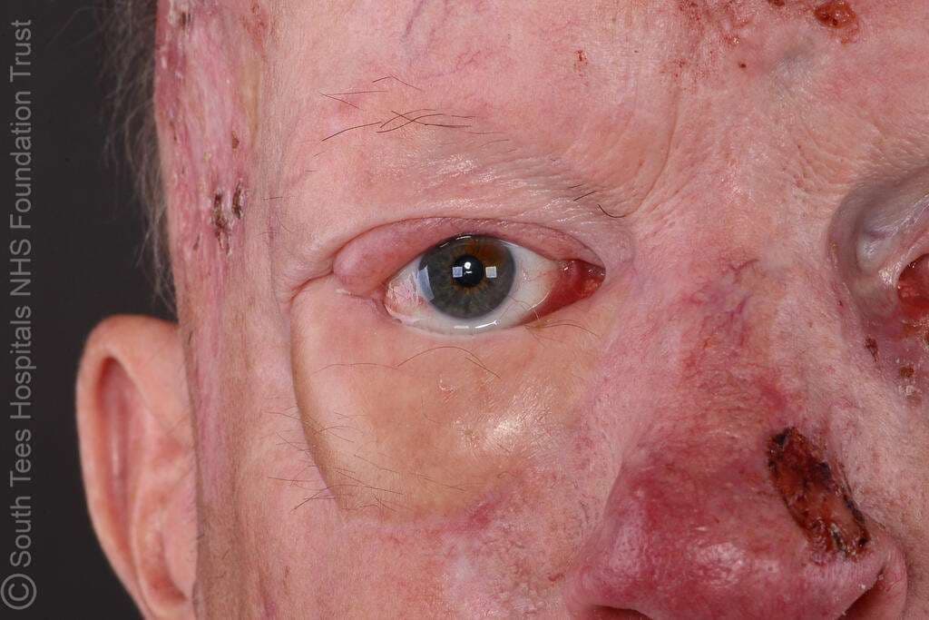
A 59-year-old male with a longstanding history of Gorlin–Goltz syndrome (Nevoid Basal Cell Carcinoma Syndrome) diagnosed in 1997 has been under multidisciplinary care involving dermatology, plastic surgery, oncology, and ophthalmology for multiple recurrent basal cell carcinomas. His medical history includes hypertension, iron deficiency anaemia secondary to diverticulosis, chronic gastritis with duodenal ulcer disease, and melanoma in situ of the right knee excised in 2016. He has undergone numerous treatments for cutaneous malignancies including repeated surgical excisions, Mohs micrographic surgery, topical therapies (5-fluorouracil and imiquimod), and reconstructive procedures. In 2012 he underwent Mohs surgery for an infiltrative basal cell carcinoma of the left lower eyelid. Over subsequent years, multiple lesions were treated across the face, temples, ears, forearm, and calf. Since April 2016 he has been managed with the Hedgehog pathway inhibitor Vismodegib with intermittent dosing. He later developed cicatricial ectropion of the right lower eyelid following prior tumour excisions, previously managed with a Hughes flap. In 2024 he underwent Mohs excision for recurrent basal cell carcinoma of the right lower eyelid, leaving a large full-thickness defect that required complex reconstruction with a radial forearm free flap. He continues long-term surveillance due to ongoing risk of tumour recurrence.

The defect includes loss of both the anterior and posterior lamellae, with involvement of the lateral canthus and medial canthal ligament, and extends to the conjunctival fornix. After confirmation of clear margins, the wound was debrided and margins freshened, removing devitalised tissue and creating a well-vascularised wound bed with exposed conjunctiva and periocular tissues, ready for definitive reconstruction.

A radial forearm free flap was designed over the radial artery territory on the volar forearm, with the skin paddle tailored to the size of the anterior lamellar defect. The radial artery and venae comitantes were dissected to obtain an adequate pedicle for microvascular anastomosis.
The palmaris longus tendon was identified and harvested with the flap to be used as a suspensory sling for eyelid support, providing structural stability similar to the tarsoligamentous system.
2

For this complex defect, a composite reconstruction was planned:
Posterior lamella: harvested hard palate graft to provide structural support and mucosal lining.
Anterior lamella: radial forearm free flap for skin coverage.
Eyelid support: palmaris longus tendon sling integrated into the flap for dynamic support.
Pre-harvest marking of the hard palate is critical to define graft dimensions while preserving the mucoperiosteum and avoiding injury to the greater palatine artery. The flap and tendon dimensions are calculated to match the defect and ensure proper eyelid contour and function post-reconstruction.
3

The radial forearm free flap was inset into the periocular defect to reconstruct the anterior lamella and provide soft-tissue coverage of the lower eyelid. The flap was carefully contoured to match the shape and size of the defect and secured to the surrounding eyelid margins using fine interrupted sutures (5-0 Monocryl and 5-0 Ethilon) to achieve precise alignment.
The palmaris longus tendon sling was positioned to support the reconstructed eyelid and was anchored to the periosteum of the medial and lateral orbital rim using 5-0 Vicryl, restoring eyelid suspension and maintaining appropriate lid position.
Following microvascular anastomosis of the radial artery to the facial artery and venous drainage to the facial vein using a 2.5-mm coupler, the flap demonstrated good perfusion, and the reconstruction was completed with layered closure.
4

This intra-operative photograph demonstrates the harvested hard palate mucoperiosteal graft, obtained from the patient’s palate for use in eyelid reconstruction. The graft provides rigid mucosal tissue with structural support, making it well suited for posterior lamella reconstruction of the lower eyelid. After careful elevation from the hard palate with preservation of surrounding mucosa, the graft is trimmed to the required size and prepared for inset into the eyelid defect. The donor site is typically managed with haemostasis and allowed to heal by secondary intention or protected with a dressing plate.
5

This intra-operative photograph demonstrates the final appearance following completion of the reconstruction of the right lower eyelid defect. The radial forearm free flap has been inset to recreate the anterior lamella, while the hard palate mucosal graft forms the posterior lamella. The palmaris longus tendon sling has been secured to the periosteum to provide structural eyelid support and restore lower eyelid suspension. The flap appears well perfused with satisfactory contour and alignment of the reconstructed eyelid. This image was taken at the end of the procedure prior to extubation.
6

Clinic photo
7

Clinic Photo - good eye closure
8

Clinic Photo
9

Clinic Photo
10
Short-Term Management (First 1–2 Weeks):
1) Flap Monitoring - Regular free flap observations (colour, capillary refill, temperature, turgor, Doppler).
2) Haemodynamic Support - Maintain systolic BP >100 mmHg, monitor urine output and ensure adequate hydration.
3) Medications - LMWH 40 mg daily for VTE prophylaxis, broad-spectrum antibiotics for 5–7 days.
4) Ocular Care - Lubricating eye drops and ointment to prevent corneal exposure. Ophthalmology review if needed.
5) Wound Care - Topical antibiotic ointment to eyelid reconstruction site, monitor radial forearm donor site and skin graft.
6) Oral Donor Site Care - Soft diet and antiseptic mouth rinses following hard palate graft harvest.
Long-Term Management:
1) Functional Assessment
2) Regular review of eyelid position, lid closure, and ocular protection.
3) Scar and Flap Monitoring - Assess flap contour, scar maturation, and need for revision procedures.
4) Donor Site Follow-up - Monitor radial forearm donor site healing and function.
5) Oncological Surveillance - Lifelong dermatologic follow-up due to risk of recurrent BCCs in Gorlin–Goltz syndrome.
6) Multidisciplinary Care - Ongoing follow-up with plastic surgery, dermatology, oncology, and ophthalmology.
How to cite this article
Miss Ketki Kaushal, Mr Oliver Bassett. Case 21 2026: Reconstructing the Challenging Eyelid: Hard Palate Graft and Radial Forearm Free Flap Reconstruction in Gorlin–Goltz Syndrome. Journal of Plastic, Breast & Reconstructive Surgery. 2026.
Open access. © 2026 The Author(s). Published by the Journal of Plastic, Breast & Reconstructive Surgery. Copyright to this case is shared between the author(s) and the journal; authors may reuse their own case for education, presentations and social media.
Keep reading
Related cases
Case 20 2026: Staged Excision and Delayed Primary Closure of a large Nodular Basal Cell Carcinoma of the Anterior Scalp
Mikkel Halborg Sørensen, Nanja Gotland Sundstrup, Christian Lyngsaa Lang
Keywords: Basal cell carcinoma, scalp reconstruction, staged excision, primary closure, scalp biomechanics. Authors: Mikkel Halborg Sørensen, MD; Nanja Gotland Sundstrup, MD; Christian Lyngsaa Lang, MD. Institution: Department of Plastic Surgery and Burns, Rigshospitalet, Copenhagen University Hos-pital, Denmark Abstract A 48-year-old woman was referred with a non-radically excised nodular basal cell carcinoma of the anterior scalp complicated by wound infection and dehiscence. Re-excision with 3 mm margins to the galea was performed under local anesthesia. Given local inflammation and uncertain margin status, delayed reconstruction was planned pending histopathological confirmation. After clear margins were verified, controlled subgaleal undermining allowed tension-reduced primary closure. This staged strategy ensured oncologic safety while preserving hair-bearing scalp and avoiding unnecessary flap reconstruction. Patient medical history A 48-year-old woman was referred after incomplete excision of a nodular basal cell carcinoma (nBCC) of the anterior scalp performed in private plastic surgery practice. Histology demonstrated tumor involvement of the lateral margins, while the deep margin was free of tumor. Postoperatively, wound rupture and infection developed. Microbiology showed sparse growth of Staphylococcus aureus, and antibiotic therapy was initiated. The patient was otherwise non-smoking and healthy, with well-treated asthma, as her only relevant comorbidity. Before and After Patient examination Clinical examination revealed a 22 × 19 mm anterior scalp defect with limited perifocal erythema and minimal purulent discharge. No regional lymphadenopathy was palpable. Pre-operative considerations Initial histopathology demonstrated nodular basal cell carcinoma with focal lateral margin involvement and a clear deep margin, without perineural invasion or other high-risk features. In accordance with current recommendations for low-risk BCC, re-excision with 3 mm c
Case 22 2026: Superior Gluteal vessel: A rarely used recipient for free flap reconstruction of lumbosacral defects: a case report.
Tushar Dutta, Arun PS, Swati Sattavan
Keywords: Superior gluteal vessel, free flap reconstruction, lumbosacral defects Authors: 1. Tushar Dutta, Consultant Plastic Surgeon; 2. Arun PS, Consultant Surgical Oncologist; 3. Swati Sattavan, Consultant Surgical Oncologist; Institute: State Cancer Institute, Guwahati, Assam, India Abstract Lumbosacral defects that are too large for local flap reconstruction should be considered for microvascular free tissue transfer. However, lack of suitable recipient vessels in the area makes the task technically difficult. Options such as thoracodorsal and inferior gluteal vessels extended with vein grafts and AV loops have been described. Each option has its own drawbacks. Despite having a short course at its origin, the superior gluteal vessel is a viable alternative. We describe here a case where the vessel was dissected out with adequate length and used as a recipient for free flap reconstruction. Patient medical history The patient was a 41-year-old lady with no known comorbidities. She presented to us with a large lumbosacral mass that had been growing for one year. Before visiting our center, she had been operated twice at other institutions and had recurrences after each occasion. During the last three months the mass had started to grow rapidly and caused ulceration. There was no pain or fever. There was no neurological involvement of lower limbs. Patient had an ECOG status 1. Before and After Patient examination A 17x 22 cm mass was noted encompassing the left part of lumbosacral region and crossing the midline to the opposite side. The superior limit of the lesion was at the L3 vertebra and inferior extent was near the coccyx. Skin ulceration was noted at the center of the mass. On palpation, the mass was warm to touch, hard in consistency and immobile. Skin was fixed and fixity was also noted to the underlying muscle. No tenderness was elicited. Surrounding possible donor sites for flap harvest showed no scars. Pre-operative considerations Preoperative biopsy co
Case 23 2026: “Minimally Invasive Finger Amputation Using a Volar ‘Toilet seat’ Flap for Invasive Squamous Cell Carcinoma”
Claes Hannibal Killerich, Nikolaj Warming
Keywords: Squamous cell carcinoma; finger amputation; volar flap; local reconstruction; traction neurectomy; hand surgery. Authors: Claes Hannibal Killerich, Nikolaj Warming Abstract A 93-year-old woman presented with a chronic non-healing dorsal finger wound initially diagnosed as actinic keratosis. Repeat biopsy revealed invasive squamous cell carcinoma with joint and bone involvement. Due to the extent of invasion, distal amputation of the third finger was required. Reconstruction was performed using a volar “toilet seat” flap under digital nerve block to minimize surgical burden. The procedure achieved clear margins, preserved stump length, and avoided donor-site morbidity. This case highlights the importance of early re-biopsy of non-healing lesions and adapting surgical strategy to patient age, function, and comorbidity. Patient medical history A 93-year-old woman presented with a wound on the dorsal aspect of the proximal phalanx of the left third finger. A primary biopsy revealed actinic keratosis, and the lesion was treated several times with curettage and electrodesiccation by a private dermatologist. Despite repeated treatments under professional supervision, the wound failed to heal.Due to the persistent non-healing nature of the lesion, a repeat biopsy was performed, confirming the diagnosis of squamous cell carcinoma (SCC). The patient was referred to the Department of Plastic Surgery, Aalborg University Hospital, where a primary excision was performed with a 7 mm surgical margin. Histopathological examination demonstrated invasive tumor growth involving both joint and bone. The defect was subsequently reconstructed using a full-thickness skin graft. Before and After Patient examination The left third finger showed a healed full-thickness skin graft on the dorsal aspect of the proximal phalanx, covering the proximal interphalangeal (PIP) joint. There was no visible residual tumor, and no palpable lymphadenopathy in the left cubital fossa or axillary re