
Case 13: Advancing Nasal Ala Reconstruction: Novel Single-Stage Bullhorn Flaps vs. Traditional Two-Stage Nasolabial Techniques
Lukas Kure-Rosenberg1, Nicco Krezdorn1, Chief1
- 1Department of Plastic & Breast Surgery, Zealand University Hospital, Denmark
Abstract
Keywords: ala nasi, nasolabial flap, infranasal flap, bullhorn modification, facial reconstruction Authors: Lukas Kure-Rosenberg, MD, Nicco Krezdorn, MD, Chief. Institution: Department of Plastic & Breast Surgery, Zealand University Hospital, Denmark Abstract Reconstructive surgery of the caudal ala nasi poses a particular challenge due to the complex anatomy and high aesthetic significance of the area. The skin in this region is thick, inelastic, and poorly mobile, and the alar contour plays a crucial role in the overall symmetry and three-dimensional shape of the nose. Additionally, local tissue availability is limited, making flap design and execution technically demanding. Two-stage procedures, which are often required in this area, can be disadvantageous as they involve prolonged treatment time, multiple interventions, and temporary aesthetic or functional compromise between stages, potentially affecting patient satisfaction. Preoperative considerations should include a detailed assessment of the defect’s size, depth, and proximity to the alar rim, as well as evaluation of nasal symmetry, skin quality, and patient expectations. It is also important to plan for structural support, such as cartilage grafts, if there is a risk of alar collapse or distortion. This report presents three cases involving patients treated for basal cell carcinoma (BCC) on the caudal part of the nasal ala, following excision with frozen section guidance. Case 1 underwent a traditional two-stage nasolabial flap and demonstrated the subsequent need for revision due to dynamic tension and functional concerns. Case 2 introduces a novel single-stage, medially based infranasal flap – a "modified half bullhorn approach" – tailored to address smaller defects in a single-stage, cosmetically discreet and functionally balanced manner. Case 3 introduces an even larger single-stage, medially based contralateral bullhorn transposition flap with advancement closure – a "modified full bullhorn appr
 BeforeAfter
Case 1, a 72-year-old female referred to the Department of Plastic Surgery, Roskilde, Sealand University Hospital, Denmark, in fall 2024, following referral by a dermatologist for surgical treatment of a lesion on the right lower alar edge. Biopsy revealed nodular basal cell carcinoma. The patient had a medical history of type 2 diabetes mellitus, asthma bronchiale, hypertension arterialis essentialis, and was an active smoker. Case 2, a 67-year-old male, was referred by a dermatologist to the Department of Plastic Surgery, Roskilde, in spring 2025 for treatment of a lesion on the lower left alar edge, with biopsy confirming nodular basal cell carcinoma. Case 3, a 82-year-old female, was referred by a dermatologist to the same department in spring 2025 for treatment of a lesion involving the infranasal region with extension into the nasal vestibule and lower left ala. Biopsy revealed nodular basal cell carcinoma. She had a history of type 2 diabetes mellitus.

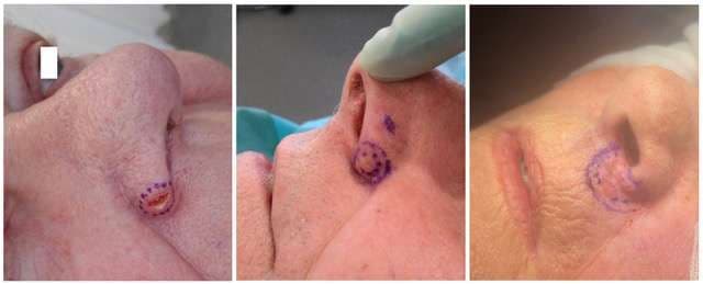
Frozen sections from 12, 3, 6, 9 o’clock and deep margins were sent for histological evaluation. After confirmation of tumor clearance, the 10 mm defect was reconstructed using a classic nasolabial flap. An inverted pexing suture (5.0 Vicryl) was placed to minimize volume at the alar groove while preserving flap perfusion. The flap was closed using inverted 5.0 Vicryl single sutures and 5.0 nylon skin sutures.
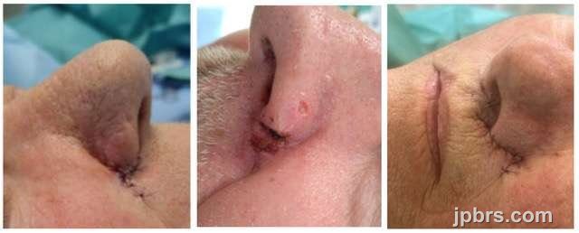
At 3-month follow-up, the patient did not report cosmetic concerns, but described nasal deviation to the contralateral side, and dynamic traction during speech and eating. Second-stage flap division was scheduled.
Images: Immediate postoperatively (left picture) demonstrated the nasolabial reconstruction and although special care was taken to minimize volume at the alar edge, the need for revision was seen at subsequent follow up (right picture).

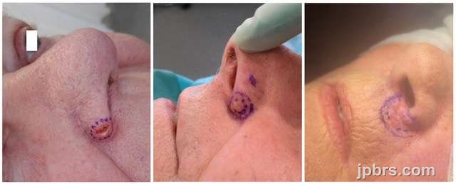
The patient was seen in an outpatient setting for second stage flap division and revision and was preoperatively marked, the more medial aspect communication with the nasal vestibule (not shown preoperatively) demonstrated a small diastasis and since the nose also shifted slightly to the left opposite of the reconstruction, this area was also raised and resected until symmetry (see image below, right picture). The patient chose to have suture removal after the second-stage revision carried out at their general practitioner, so no further follow-up images are available. The patient was postoperatively informed to request further consult if needed.
Images: Preoperative markings of the abundant flap tissue to be excised (left picture). The flap was also lifted and reattached at the medial aspect communicating with the vestibule, to address shifting of the nose to the left. Immediate post operative of the second stage (medial pictures and right picture).
2

Frozen sections from 12, 3, 6, 9 o’clock and deep margins were negative. An additional 2mm medial excision on the alar was also confirmed tumor-free.
The excised defect measuring 11mm in diameter and initial markings (left picture). The proposed flap is marked in green (second picture left), the excised burrow triangle is marked with red, and the subsequent advancement is marked in yellow. Red lines identify incision lines (middle picture). Closure of the primary defect with flap transposition, and the following secondary defect marked in red (second picture right). The infranasal area transposition of the advancement marked in yellow (right picture).
3

The flap and secondary advancement were sutured with inverted 5.0 vicryl single suture and skin closure using 5.0 nylon single sutures.
Images immediate postoperative, demonstrate good cosmesis and perfusion. Notice there is no traction in the upper lip. The area on the alar where supplementary histopathology had been preformed, was dressed for conservative healing. No bleeding or complications was observed. Sutures were planned for removal at following 7-day visit.
4

A small superficial wound was noted medially, suitable for conservative healing. The patient was highly satisfied with the aesthetic and functional result and declined further follow-up.
5

Initial markings of tumor only (left picture), not addressing the preoperative visible area of tumor on the right alar and nasal vestibule (right picture). The full involvement of the nasal vestibule is not marked, since the alar was surgically opened before final markings was performed.
6

Frozen sections from 12 (vestibular floor), 3 (lateral ala), 6 (supralabial), 9 (columella), and deep margin confirmed clear margins. The final defect measured 26 × 13 mm, involving 5 × 7 mm of lower vestibular floor. Lower alar cartilages and fibro-fatty tissue lateral were left intact (Left picture). Tissue mobility and lip position was assessed before the reconstruction was started (Left middle picture).
A back-cut was preformed in the left nasolabial crease and a small perialar burrow triangle was made in the skin (only) of alar grove (Center, center right picture) to adresse the larger primary defect on the left side before further reconstruction, aiming at maintaining symmetry and adding tissue to the unsupported left lower alar.
The contralateral infranasal flap was raised in a 90° transposition manner on a medial base. Its width (5 mm) matched the vestibular defect. The flap was marked at 7mm, equal to the length of the vestibular defect after advancement of the area. The symmetrizing excision of abundant skin after the 7mm mark was raised just below the skin, where the first 7 mm of the pedicle was raised on subcutaneous plane (Right picture)
7

The two sides of the infranasal regio was advance for final closure.
Flap fixation used 5.0 Monosyn rapide (vestibule) and 5.0 Vicryl for deep sutures, with 5.0 nylon continuous skin closure. Prophylactic dicloxacillin was prescribed due to DM2 and nasal cavity involvement.The reconstruction and flap demonstrated good cosmesis and good vascular perfusion immediate postoperative with no bleeding or complications. Surgery occurred on the case submission deadline date.
Images: The flap immediate postoperatively. Notice almost equal lifting of the upper lip. is no traction in the upper lip. Although the base of the alar edge was secured at its base before the nasolabial advancement was sutured, there is a discreet downward shift of the alar position on the left side. The patient was pleased with the initial outcome of the reconstruction.
8
Reconstruction of the caudal nasal ala poses significant challenges due to mobility, contour complexity, and aesthetic sensitivity. While nasolabial flaps remain the gold standard, they often require staging. Modified infranasal (bullhorn) flaps offer a cosmetically discreet, single-stage alternative for suitable small to moderate defects, improving both patient satisfaction, outcomes and surgical efficiency.
How to cite this article
Lukas Kure-Rosenberg, Nicco Krezdorn, Chief. Case 13: Advancing Nasal Ala Reconstruction: Novel Single-Stage Bullhorn Flaps vs. Traditional Two-Stage Nasolabial Techniques. Journal of Plastic, Breast & Reconstructive Surgery. 2025.
Open access. © 2025 The Author(s). Published by the Journal of Plastic, Breast & Reconstructive Surgery. Copyright to this case is shared between the author(s) and the journal; authors may reuse their own case for education, presentations and social media.
Keep reading
Related cases
Case 17: Immediate Proximal Row Carpectomy for combined Scaphoid and Lunate Dislocation Without Fracture
Jorge Jimenez Lopez, Anastasia Boptsi, Konstantinos Kalousis
Keywords: Hand surgery; wrist; carpectomy; scaphoid; lunate dislocation Authors: Jorge Jimenez Lopez, MD; Anastasia Boptsi, MD; Konstantinos Kalousis, MD Institution: Universitätsklinikum Schleswig-Holstein Lübeck Abstract Carpal dislocation fractures are uncommon injuries, most frequently associated with high-energy trauma. Combined carpal dislocations without associated bone fractures are even rarer. Delayed treatment can have serious consequences. The case presented here involves an 86-year-old man with dislocation of the scaphoid and lunate bones, as well as acute carpal tunnel syndrome resulting from the injury. PRC is mostly a salvage operation but given the severity of ligamentous disruption, the need to restore the patient's mobility and also the fact, that scaphoid was irreducible, a proximal row carpectomy (PRC) was performed to preserve wrist range of motion (ROM). Patient medical history The 86-year-old patient crashed the car into the wall of his garage at a speed of 30 km/h. As a result, the patient presented a dislocation of the lunate bone and the scaphoid. The patient lived alone and was completely independent. Regarding other conditions, he suffered from arterial hypertension and prostate cancer. Before and After Patient examination There was a visible deformity at the right wrist with bruising, and swelling. Additionally, a positive Hoffman-Tinel sign was observed with paresthesia in the median nerve area. A radiological examination of the hand was then performed, which revealed a dislocation of the scaphoid and lunate bones. No bone fractures were detected. Pre-operative considerations Given the severity of ligamentous disruption and the need to restore the patient’s mobility a proximal row carpectomy (PRC) was performed to preserve wrist range of motion (ROM). 1 Surgical Dorsal Exposure The surgical procedure involved exposing and splitting the 4th and 3rd extensor tendon compartments to facilitate access. The extensor pollicis longus (EPL) tend
Case 16: Utilizing the reverse radial forearm flap for burn wound management of the hypothenar part of hand
David Salim, MD, Msc
Keywords: Reverse radial forearm flap, Burn wound management, Hand surgery Authors: David Salim, MD and Taiba Alrasheed, MD, FRCSC, Msc. Institution: Department of Plastic- & Breast Surgery , Zealand University Hospital Roskilde, Denmark Abstract A 55-year-old man had sought and was admitted to the emergency department one week after sustaining a superficial second degree burn wound on the hypothenar region of the right hand after accidentally placing it on a hot stove. At first sight, the burn wound seemed quite superficial and conservative healing and bandaging was initially deemed sufficient with the aim of spontaneous healing. One week after the initial assessment in the emergency department, the patient was seen for a routine clinical control of the wound. At this consultation, the patient scored 4/4 positive Kanavel’s signs and flexor tenosynovitis was suspected. Afterwards immediate wound revision of the right hypothenar was conducted. During wound revision, considerable amounts pus was noted and furthermore the musculus abductor digiti minimi was avital and excised. Suitable wound dressing was applied, pus was sent for culture and sensitivity testing and the patient was gives appropriate peroral antibiotics. After numerous wound revisions and debridement, control of the infection at the orthopedic surgery department was achieved. The patient was then referred to the plastic surgery department for planning of soft tissue coverage with a reverse radial forearm flap. Patient medical history The patient was known to have substantial unregulated diabetes mellitus type two, but no other known medical comorbidities. Before and After Patient examination The patient was seen in an outpatient setting in the plastic surgery department for planning of surgery. The defect was located to the whole hypothenar region of the right hand. Substantial soft tissue loss was noted and the 3-5th flexor tendons were exposed. Pre-operative considerations A reverse radial forearm
Case 15: Closure of a complex defect after a dog bite wound of the middle third of the face
Dr. Guillermo Alvarez-Sanchez, Ignacio Lugo-Beltran
Keywords: Cheek reconstruction, face, cheek, forearm, free flap, Neurorrhaphy , radial forearm flap Authors: Dr. Guillermo Alvarez-Sanchez, MD ; Ignacio Lugo-Beltran, MD, Chief, Dr. Haro-Cruz Jorge Said, MD. Institution: Plastic and Reconstructive Surgery Division of Microsurgery, Hospital Angeles Metropolitano, Cuauhtémoc, Mexico. Abstract A 52 year old female who presents to the emergency room after receiving a dog bite in her right face, the defect is managed with a free radial forearm flap. Patient medical history 52 year old female, without past medical history, presented to the emergency room, referring she was attacked by her dog at her house, suffering a bite on her right cheek, with bleeding and loss of skin Before and After 1 Before photo - Arrival at the ER Facial asymmetry, with a complex wound on the right cheek, with skin loss, a defect approximately 10 x 9 cm, irregular borders, with muscle exposure, visualizing blood vessels and section of the buccal and zygomatic terminal nerve branches, active mild bleeding. Intact parotid gland and Stensen duct. Laceration of the zygomatic ligament. Difficulty mobilizing of the zygomatic and buccal branches muscle groups. Patient examination Facial asymmetry, with a complex wound on the right cheek, with skin loss, a defect approximately 10 x 9 cm, irregular borders, with muscle exposure, visualizing blood vessels and section of the buccal and zygomatic terminal nerve branches, active mild bleeding. Intact parotid gland and Stensen duct. Laceration of the zygomatic ligament. Difficulty mobilizing of the zygomatic and buccal branches muscle groups. Pre-operative considerations Given the circumstance of tissue loss, active bleeding and section of the nerve branches, also being a contaminated wound, we decided to go to the operating room immediately Due to tissue loss dimensions, we planned a radial free flap for tissue coverage 2 Debridement and assessing damages we cleansed, remodeled the irregular borders and clos