
Case 12: Flower Flap - A Novel Papilla Reconstruction Technique Using Local Flaps and Skin Graft
Anna Louise Norling1, Nanja Gotland Sundstrup1, Matilda Svenning Hunt1
- 1Department of Plastic Surgery and Burns, Rigshospitalet, Copenhagen University Hospital, Denmark
Abstract
Keywords: Flower Flap, Papilla reconstruction, nipple reconstruction, breast revision, full-thickness skin graft, dog ear correction Authors: Anna Louise Norling, MD, Nanja Gotland Sundstrup, MD, Matilda Svenning Hunt, MD and Pia Cajsa Leth Andersen, MD. Institution: Department of Plastic Surgery and Burns, Rigshospitalet, Copenhagen University Hospital, Denmark Abstract A 47-year-old woman underwent bilateral papillae reconstruction using a novel reconstruction technique involving four local flaps and a full-thickness skin graft including subcutaneous tissue from excised dog ears, due to scar correction, following a DIEP flap breast reconstruction. This technique preserved breast projection, avoided additional scarring, and improved breast contour. Initial epidermolysis occurred, as expected with a thick graft, but the grafts survived. At one month follow-up, the papillae showed resolving discoloration. This simple and reliable method facilitates simultaneous nipple reconstruction and aesthetic breast contouring with minimal donor-site morbidity. Patient medical history A 47-year-old woman with a history of DCIS in the left breast and a family history of breast cancer (BRCA-negative) initially underwent bilateral subcutaneous mastectomy with NAC removal and implant-based reconstruction in 2021. Due to dissatisfaction with the result, she had bilateral DIEP flap reconstruction in May 2023. In July 2024, a revision was performed to improve the breast shape, including correction of dog ears and redundant skin on the DIEP flaps, as well as lateral liposuction. At the 3-month follow-up, the breasts remained laterally full with a square appearance. A new revision was planned to refine the contour and simultaneously reconstruct the nipple using a novel technique combining four small local flaps and a full-thickness skin graft with subcutaneous tissue. Before and After Patient examination The patient was a well-preserved woman with good-quality skin. Clinical examination r
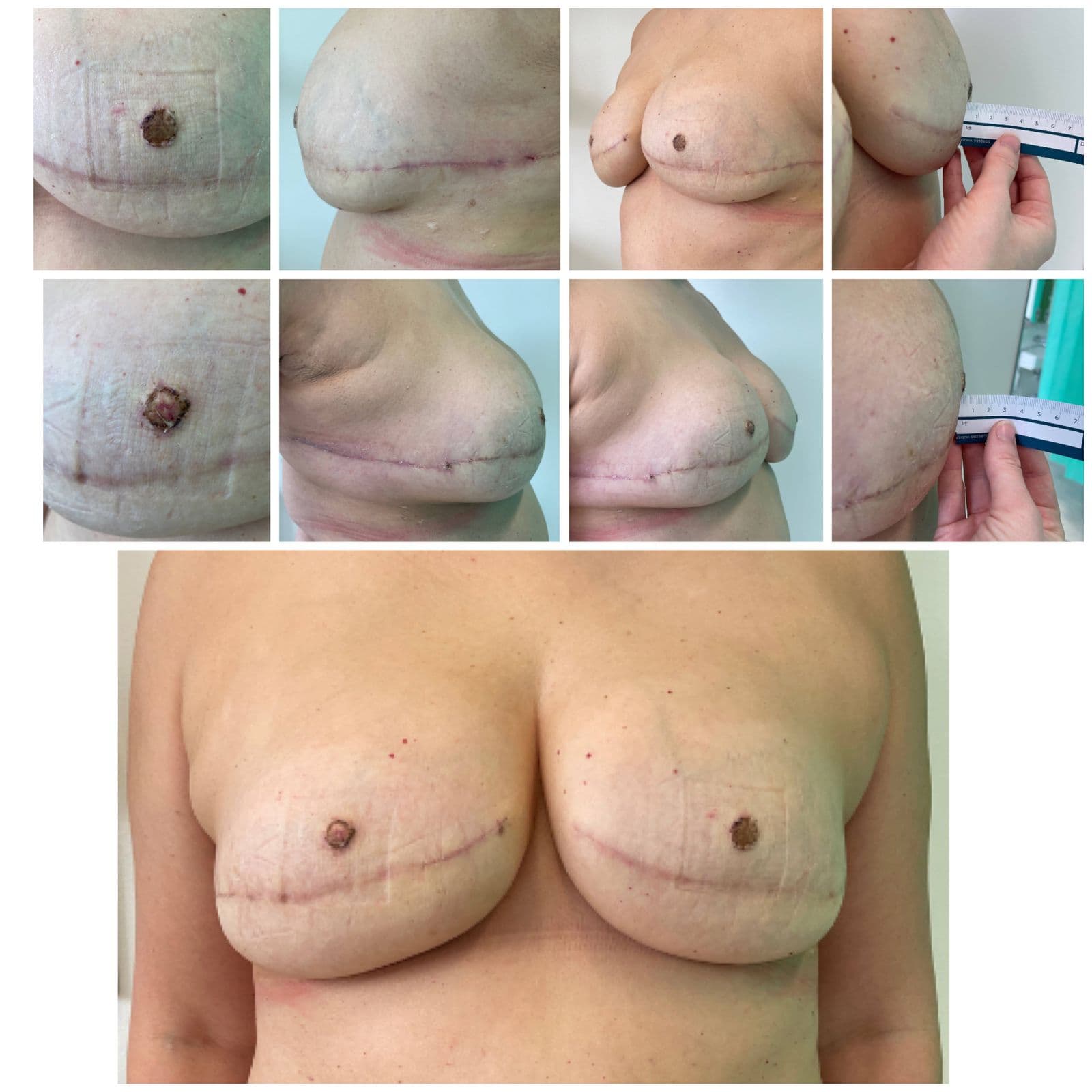
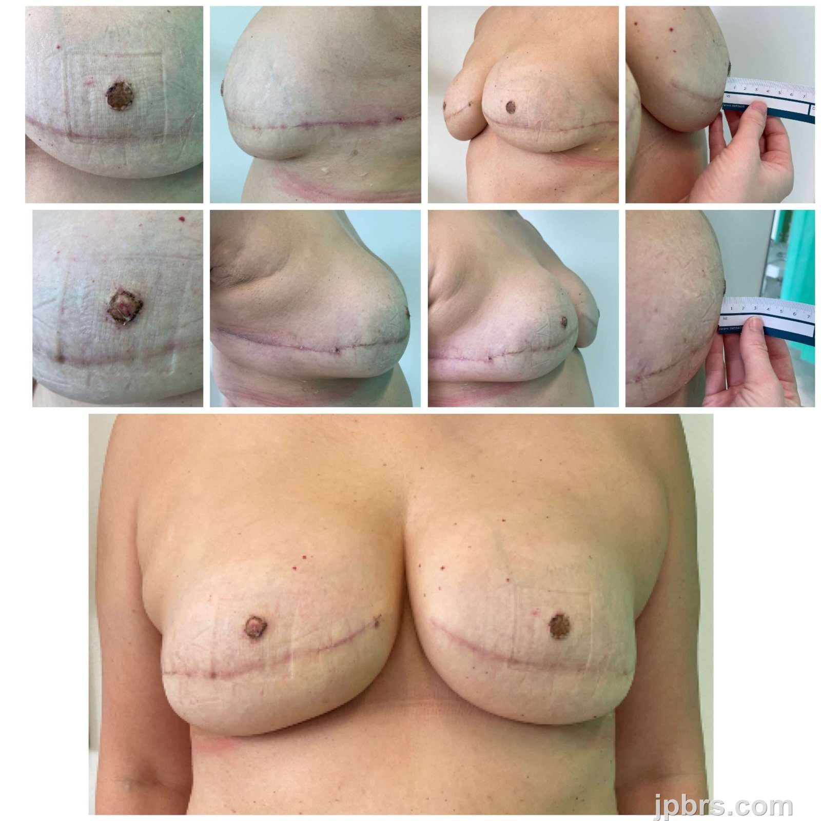
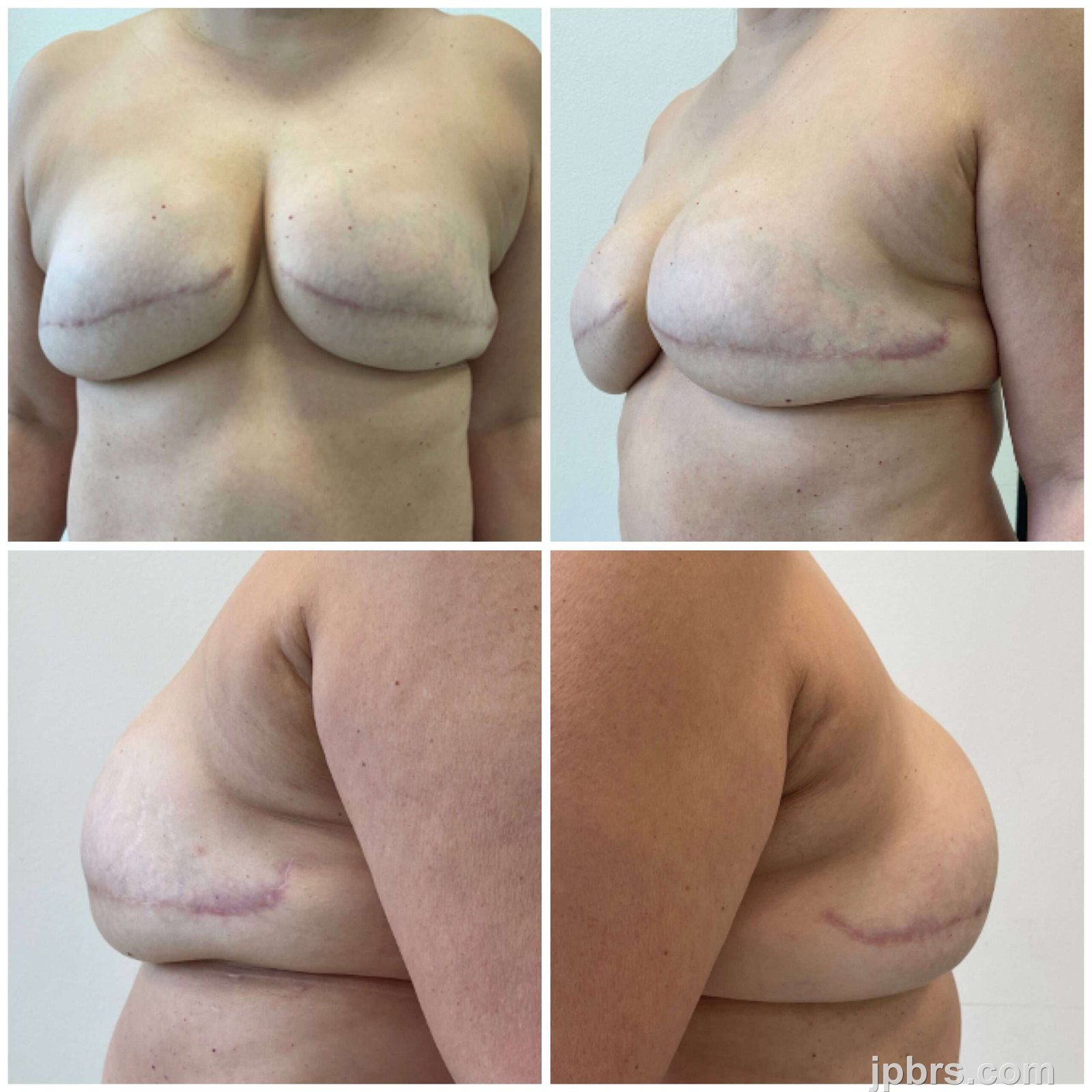 BeforeAfter
A 47-year-old woman with a history of DCIS in the left breast and a family history of breast cancer (BRCA-negative) initially underwent bilateral subcutaneous mastectomy with NAC removal and implant-based reconstruction in 2021. Due to dissatisfaction with the result, she had bilateral DIEP flap reconstruction in May 2023. In July 2024, a revision was performed to improve the breast shape, including correction of dog ears and redundant skin on the DIEP flaps, as well as lateral liposuction. At the 3-month follow-up, the breasts remained laterally full with a square appearance. A new revision was planned to refine the contour and simultaneously reconstruct the nipple using a novel technique combining four small local flaps and a full-thickness skin graft with subcutaneous tissue.

Incision lines were drawn to address lateral fullness and dog ears, shaping the breasts accordingly.

Papilla prosthetics were tried on in front of a mirror with the patient to determine optimal nipple placement based on the planned breast contour.
2

The site for papillae reconstruction was marked.
3

Local anesthesia was administered for dog ear excision.
4

Dog ears were excised and closed up with primary sutures using Vicryl 3-0 for the subdermis and Caprosyn 4-0 intradermally.
5

The excised skin and subcutaneous tissue(the medial dog ear and the lateral dog ear on the left breast) were collected.
6

Portions of the excised tissue, typically discarded, were used to create new papillae.
The grafts were shaped into 1 cm diameter circles, with excess subcutaneous tissue trimmed while retaining some to increase height of the papillae.
7

The final grafts consisted of full-thickness skin with subcutaneous tissue.
8

The incision lines for the four small triangular local flaps were marked.
9

The flaps were raised with underlying subcutaneous tissue, creating a central wound bed for graft placement. The raised local flaps were assessed for adequate mobility.
10

The grafts were sutured in place using Prolene 5-0. Sutures were placed at the four corners of the local flaps and the corresponding areas on the graft, with additional sutures along the grooves between the flaps.
11

Immediate postoperative evaluation showed the height and appearance of the reconstructed papillae.
12

A compression dressing of Jelonet, gauze, and Micropore was applied, with no need for sutures.
13

On postoperative day 7 (POD 7), the dressing was removed. As expected, the graft appeared dark with epidermolysis, typical for a skin graft that includes subcutaneous tissue.
14

By POD 15, the grafts were clearing, with graft survival evident.
15

On POD 31, the papillae remained mildly discolored but continued to improve with expected normalization anticipated within the following month.
16
How to cite this article
Anna Louise Norling, Nanja Gotland Sundstrup, Matilda Svenning Hunt. Case 12: Flower Flap - A Novel Papilla Reconstruction Technique Using Local Flaps and Skin Graft. Journal of Plastic, Breast & Reconstructive Surgery. 2025.
Open access. © 2025 The Author(s). Published by the Journal of Plastic, Breast & Reconstructive Surgery. Copyright to this case is shared between the author(s) and the journal; authors may reuse their own case for education, presentations and social media.
Keep reading
Related cases
Case 17: Immediate Proximal Row Carpectomy for combined Scaphoid and Lunate Dislocation Without Fracture
Jorge Jimenez Lopez, Anastasia Boptsi, Konstantinos Kalousis
Keywords: Hand surgery; wrist; carpectomy; scaphoid; lunate dislocation Authors: Jorge Jimenez Lopez, MD; Anastasia Boptsi, MD; Konstantinos Kalousis, MD Institution: Universitätsklinikum Schleswig-Holstein Lübeck Abstract Carpal dislocation fractures are uncommon injuries, most frequently associated with high-energy trauma. Combined carpal dislocations without associated bone fractures are even rarer. Delayed treatment can have serious consequences. The case presented here involves an 86-year-old man with dislocation of the scaphoid and lunate bones, as well as acute carpal tunnel syndrome resulting from the injury. PRC is mostly a salvage operation but given the severity of ligamentous disruption, the need to restore the patient's mobility and also the fact, that scaphoid was irreducible, a proximal row carpectomy (PRC) was performed to preserve wrist range of motion (ROM). Patient medical history The 86-year-old patient crashed the car into the wall of his garage at a speed of 30 km/h. As a result, the patient presented a dislocation of the lunate bone and the scaphoid. The patient lived alone and was completely independent. Regarding other conditions, he suffered from arterial hypertension and prostate cancer. Before and After Patient examination There was a visible deformity at the right wrist with bruising, and swelling. Additionally, a positive Hoffman-Tinel sign was observed with paresthesia in the median nerve area. A radiological examination of the hand was then performed, which revealed a dislocation of the scaphoid and lunate bones. No bone fractures were detected. Pre-operative considerations Given the severity of ligamentous disruption and the need to restore the patient’s mobility a proximal row carpectomy (PRC) was performed to preserve wrist range of motion (ROM). 1 Surgical Dorsal Exposure The surgical procedure involved exposing and splitting the 4th and 3rd extensor tendon compartments to facilitate access. The extensor pollicis longus (EPL) tend
Case 16: Utilizing the reverse radial forearm flap for burn wound management of the hypothenar part of hand
David Salim, MD, Msc
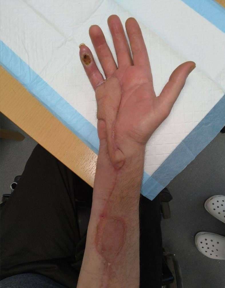
Keywords: Reverse radial forearm flap, Burn wound management, Hand surgery Authors: David Salim, MD and Taiba Alrasheed, MD, FRCSC, Msc. Institution: Department of Plastic- & Breast Surgery , Zealand University Hospital Roskilde, Denmark Abstract A 55-year-old man had sought and was admitted to the emergency department one week after sustaining a superficial second degree burn wound on the hypothenar region of the right hand after accidentally placing it on a hot stove. At first sight, the burn wound seemed quite superficial and conservative healing and bandaging was initially deemed sufficient with the aim of spontaneous healing. One week after the initial assessment in the emergency department, the patient was seen for a routine clinical control of the wound. At this consultation, the patient scored 4/4 positive Kanavel’s signs and flexor tenosynovitis was suspected. Afterwards immediate wound revision of the right hypothenar was conducted. During wound revision, considerable amounts pus was noted and furthermore the musculus abductor digiti minimi was avital and excised. Suitable wound dressing was applied, pus was sent for culture and sensitivity testing and the patient was gives appropriate peroral antibiotics. After numerous wound revisions and debridement, control of the infection at the orthopedic surgery department was achieved. The patient was then referred to the plastic surgery department for planning of soft tissue coverage with a reverse radial forearm flap. Patient medical history The patient was known to have substantial unregulated diabetes mellitus type two, but no other known medical comorbidities. Before and After Patient examination The patient was seen in an outpatient setting in the plastic surgery department for planning of surgery. The defect was located to the whole hypothenar region of the right hand. Substantial soft tissue loss was noted and the 3-5th flexor tendons were exposed. Pre-operative considerations A reverse radial forearm
Case 15: Closure of a complex defect after a dog bite wound of the middle third of the face
Dr. Guillermo Alvarez-Sanchez, Ignacio Lugo-Beltran
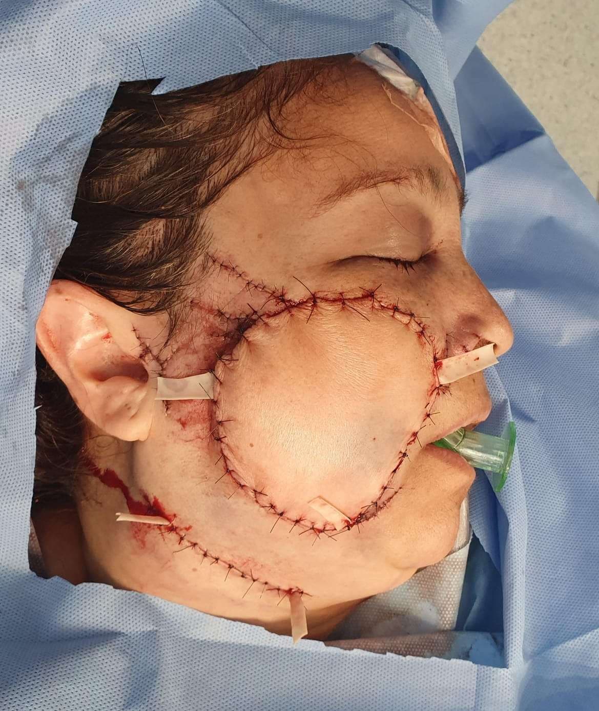
Keywords: Cheek reconstruction, face, cheek, forearm, free flap, Neurorrhaphy , radial forearm flap Authors: Dr. Guillermo Alvarez-Sanchez, MD ; Ignacio Lugo-Beltran, MD, Chief, Dr. Haro-Cruz Jorge Said, MD. Institution: Plastic and Reconstructive Surgery Division of Microsurgery, Hospital Angeles Metropolitano, Cuauhtémoc, Mexico. Abstract A 52 year old female who presents to the emergency room after receiving a dog bite in her right face, the defect is managed with a free radial forearm flap. Patient medical history 52 year old female, without past medical history, presented to the emergency room, referring she was attacked by her dog at her house, suffering a bite on her right cheek, with bleeding and loss of skin Before and After 1 Before photo - Arrival at the ER Facial asymmetry, with a complex wound on the right cheek, with skin loss, a defect approximately 10 x 9 cm, irregular borders, with muscle exposure, visualizing blood vessels and section of the buccal and zygomatic terminal nerve branches, active mild bleeding. Intact parotid gland and Stensen duct. Laceration of the zygomatic ligament. Difficulty mobilizing of the zygomatic and buccal branches muscle groups. Patient examination Facial asymmetry, with a complex wound on the right cheek, with skin loss, a defect approximately 10 x 9 cm, irregular borders, with muscle exposure, visualizing blood vessels and section of the buccal and zygomatic terminal nerve branches, active mild bleeding. Intact parotid gland and Stensen duct. Laceration of the zygomatic ligament. Difficulty mobilizing of the zygomatic and buccal branches muscle groups. Pre-operative considerations Given the circumstance of tissue loss, active bleeding and section of the nerve branches, also being a contaminated wound, we decided to go to the operating room immediately Due to tissue loss dimensions, we planned a radial free flap for tissue coverage 2 Debridement and assessing damages we cleansed, remodeled the irregular borders and clos