
Case 9: Adipofascial Turnover Flap for Reconstruction of a Dorsal Finger Defect Following SCC Excision
Lukas Kure-Rosenberg1, MD Nicco Krezdorn1, Chief1
- 1Department of Plastic Surgery, Zealand University Hospital, Denmark
Abstract
Keywords: Squamous cell carcinoma, adipofascial, turnover flap, finger, defect, reconstruction Authors: Lukas Kure-Rosenberg, MD Magnus Balslev Avnstorp, MD Nicco Krezdorn, MD, Chief. Institution: Department of Plastic Surgery, Zealand University Hospital, Denmark Abstract Reconstruction of finger defects presents unique challenges, particularly in those with complex anatomical involvement. We present a case involving an immunosuppressed patient with a larger late diagnosed SCC, the excision of- and subsequent reconstruction- utilizing an adipofascial turnover flap (APTF), that offers a reliable and versatile option for addressing these challenges, while preserving hand function and aesthetics. A 74-year-old male with a history of kidney transplantation and long-standing immunosuppressive therapy presented with a highly differentiated SCC on the dorsum of the right fourth finger. The lesion, measuring 40 × 35 mm, extended to the proximal and distal interphalangeal joints. Surgical management included two stages: tumor excision with clear margins, followed by secondary reconstruction utilizing an APTF combined with a split-thickness skin graft (STSG). Preoperative and perioperative Doppler assessments guided flap design and ensured adequate vascularity. The APTF provided robust soft-tissue coverage of the exposed extensor tendon, with adequate perfusion confirmed by Doppler. The STSG demonstrated partial take but effectively covered the underlying APTF. Postoperative assessments were followed 30, 48, and 106 days postoperatively. Range of motion (ROM) showed postoperatively some limitations in the affected finger, particularly in the PIP and DIP joints, but functionality was aimed preserved with ongoing therapy. We demonstrate in this clinical case, how APTF is an effective and versatile reconstructive option for complex dorsal finger defects. Its robust vascular supply, adaptability, and minimal donor site morbidity make it particularly advantageous in immunosuppres
Cavadas, P. C., Landin, L., & Ibáñez, J. F. (2008). Adipofascial Turnover Flap for Coverage of Dorsal Finger Defects.Moojen, D. J., et al. (2007). Treatment of Tendon and Bone Defects in the Hand Using Local Flaps.Matsui, Y., et al. (2019). Comparative Outcomes of Perforator Flaps in Hand Reconstruction.Sokolich, J. C., & Lin, C. H. (2020). Soft-Tissue Reconstruction of the Hand: Flaps and Techniques.
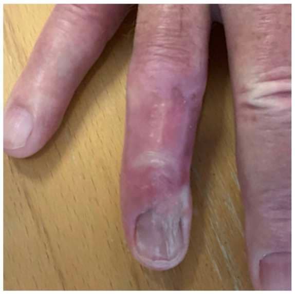
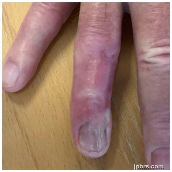 BeforeAfter
A 74-year-old male was referred to the Department of Plastic-Surgery, Roskilde, Zealand University Hospital, Denmark, fall 2024 by private plastic surgery for treatment of a lesion of the right hands fourth finger. The patient had a history of kidney transplantation and was on immunosuppressive therapy, followed regularly by the dermatology department for skin checkups. The patient had reported a persistent lesion on the dorsum of the right fourth finger for the last 4–5 years. Previously, clinically interpreted as a Verruca Vulgaris and had been recommended over-the-counter treatments without improvement. Medical records indicated cryotherapy treatments since 2018, which were also ineffective. Following establishment of squamous cell carcinoma (SCC) diagnosis via biopsy, it was deeming necessary for hospital-based treatment.

A digital nerve block was administered using 5 mL of local anesthesia (LA) without adrenaline, targeting all nerve supplies to the right fourth finger, just distal to the metacarpophalangeal joint (MTP). The surgical site was cleansed with an antiseptic solution to achieve sterile conditions. A sterile finger tourniquet was fashioned from a cut surgical glove to maintain hemostasis during the procedure.

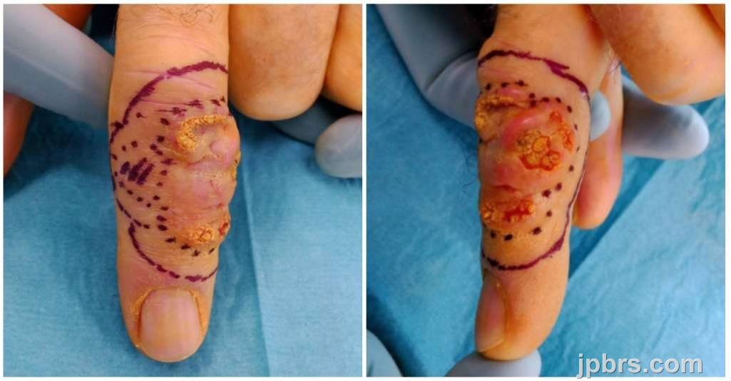
Timeout and starting from the ulnar side, incision and dissection were performed with careful visualization and preservation of nerves and vessels, proceeding to the extensor peritendon, which was exposed after tumor elevation (Figure, left). The peritendon was found to be intact, with no evidence of macroscopic tumor infiltration (Figure, center). Dissection continued within this plane toward the radial side, extending across the proximal interphalangeal (PIP) and distal interphalangeal (DIP) joint capsules. On the radial side, the dorsal digital nerve and artery were assessed adherent to the tumor and resected with the tumor specimen, which was orientation-marked and submitted for expedited hematoxylin and eosin (H&E) pathology evaluation. The macroscopically clear defect (Figure, right) was dressed in Jelonet, gauze, and a finger bandage. Follow-up and control were planned accordingly.
2

The patient was evaluated for wound care in the outpatient clinic 5 days after the first stage surgery. No signs of infection were observed, and the patient reported minimal pain, which was managed effectively with over-the-counter analgesics. Sensory deficits in radial, dorsal, the distal part of the finger was noticed as expected.
3

Eleven days after tumor excision, radical tumor removal was confirmed microscopically. Twenty days post-excision, secondary stage with closure of the defect, now measuring 40 × 40 mm, was performed.
During operative preparation, dressing removal and cleansing of the defect revealed degeneration and weakening of the extensor tendon in the distal part of the defect, resulting in partial mallet finger deformity. This was addressed during the procedure. Preoperative Doppler examination identified signal from the dorsal aspect of a. digitalis palmaris propria proximal to the defect, including signal dorsally, confirming adequate perforator flow for reconstruction.
Under sterile conditions and local anesthesia as previously, but without tourniquet, the procedure was performed following time-out. The extensor tendon was repaired using a 4-0 Prolene Kessler suture technique, restoring the tendon to its normal position.
An incision was made over the radial-dorsal aspect of the skin proximal to the defect, extending distally over the dorsum of the metacarpophalangeal joint (MTP). The skin flap was elevated with respect for the underlying structures on an ulnar base (Figure, left).
Perioperative Doppler confirmed perforator flow from the dorsal segment of the a. digitalis palmaris propria at A7, with the strongest signal originating from the radial side perforator. The adipofascial layer was mobilized from the ulnar side toward the proximal part and radially, with regards to preserving the perforator. The flap was divided longitudinally to ensure complete coverage of the tendon across the defect (Figure center) and was fixed distally with 4-0 Vicryl simple inverted sutures.
A 2.5 × 5 cm split-thickness skin graft (STSG) was harvested from the right cubit, with primary closure of the donor site. The graft was manually meshed and secured over the APFT using 4-0 Nylon interrupted sutures, ensuring preservation of the vascular supply to the underlying flap (Figure right). The wound was dressed with Jelonet, foam, a custom splint, and a finger bandage. Postoperatively, the patient was scheduled for bolus dressing removal in the outpatient clinic seven days after surgery.
4

The patient was evaluated in the outpatient clinic seven days after the second stage surgery for dressing removal. Moderate to significant swelling of the finger was observed, with normal vascular conditions distal to the surgical site. The skin graft appeared fragile with only partial take (Figure, top left), but provided adequate coverage of the APFT (which itself provided cover to the extensor tendon). In consultation with the patient, a decision was made to proceed with conservative healing. The patient was referred to ergonomical therapy and the reconstruction was subsequently evaluated at 30 days (Figure, top right), 48 days (Figure, bottom left), and 106 days (Figure, bottom right) postoperatively, demonstrating progressive improvement.
5
The patient was concurrently referred to and followed by occupational therapy during wound healing follow-up. After achieving satisfactory healing, the patient was discharged from the department of Plastic Surgery and continued regular dermatological follow-up for skin surveillance.
Once healing was complete after around 120 days, a silicone dressing was applied by occupational therapy to prevent hypertrophic scar formation. The patient exhibited persistent sensory deficits in the distal dorsal radial side of the finger but engaged in sensory training using tools such as arthro-rollers, hand trainers, and Dr. Winkler exercises. While the patient was encouraged to perform active, non-weight-bearing blocking exercises, these were not consistently practiced.
Before referral to municipal physiotherapy, the following range of motion (ROM) measurements were documented for the right fourth finger:
The patient’s ROM demonstrated limitations upon discharged. However, ongoing therapy and exercises were recommended to improve functionality further and reduce stiffness.
How to cite this article
Lukas Kure-Rosenberg, MD Nicco Krezdorn, Chief. Case 9: Adipofascial Turnover Flap for Reconstruction of a Dorsal Finger Defect Following SCC Excision. Journal of Plastic, Breast & Reconstructive Surgery. 2025.
Open access. © 2025 The Author(s). Published by the Journal of Plastic, Breast & Reconstructive Surgery. Copyright to this case is shared between the author(s) and the journal; authors may reuse their own case for education, presentations and social media.
Keep reading
Related cases
Case 17: Immediate Proximal Row Carpectomy for combined Scaphoid and Lunate Dislocation Without Fracture
Jorge Jimenez Lopez, Anastasia Boptsi, Konstantinos Kalousis
Keywords: Hand surgery; wrist; carpectomy; scaphoid; lunate dislocation Authors: Jorge Jimenez Lopez, MD; Anastasia Boptsi, MD; Konstantinos Kalousis, MD Institution: Universitätsklinikum Schleswig-Holstein Lübeck Abstract Carpal dislocation fractures are uncommon injuries, most frequently associated with high-energy trauma. Combined carpal dislocations without associated bone fractures are even rarer. Delayed treatment can have serious consequences. The case presented here involves an 86-year-old man with dislocation of the scaphoid and lunate bones, as well as acute carpal tunnel syndrome resulting from the injury. PRC is mostly a salvage operation but given the severity of ligamentous disruption, the need to restore the patient's mobility and also the fact, that scaphoid was irreducible, a proximal row carpectomy (PRC) was performed to preserve wrist range of motion (ROM). Patient medical history The 86-year-old patient crashed the car into the wall of his garage at a speed of 30 km/h. As a result, the patient presented a dislocation of the lunate bone and the scaphoid. The patient lived alone and was completely independent. Regarding other conditions, he suffered from arterial hypertension and prostate cancer. Before and After Patient examination There was a visible deformity at the right wrist with bruising, and swelling. Additionally, a positive Hoffman-Tinel sign was observed with paresthesia in the median nerve area. A radiological examination of the hand was then performed, which revealed a dislocation of the scaphoid and lunate bones. No bone fractures were detected. Pre-operative considerations Given the severity of ligamentous disruption and the need to restore the patient’s mobility a proximal row carpectomy (PRC) was performed to preserve wrist range of motion (ROM). 1 Surgical Dorsal Exposure The surgical procedure involved exposing and splitting the 4th and 3rd extensor tendon compartments to facilitate access. The extensor pollicis longus (EPL) tend
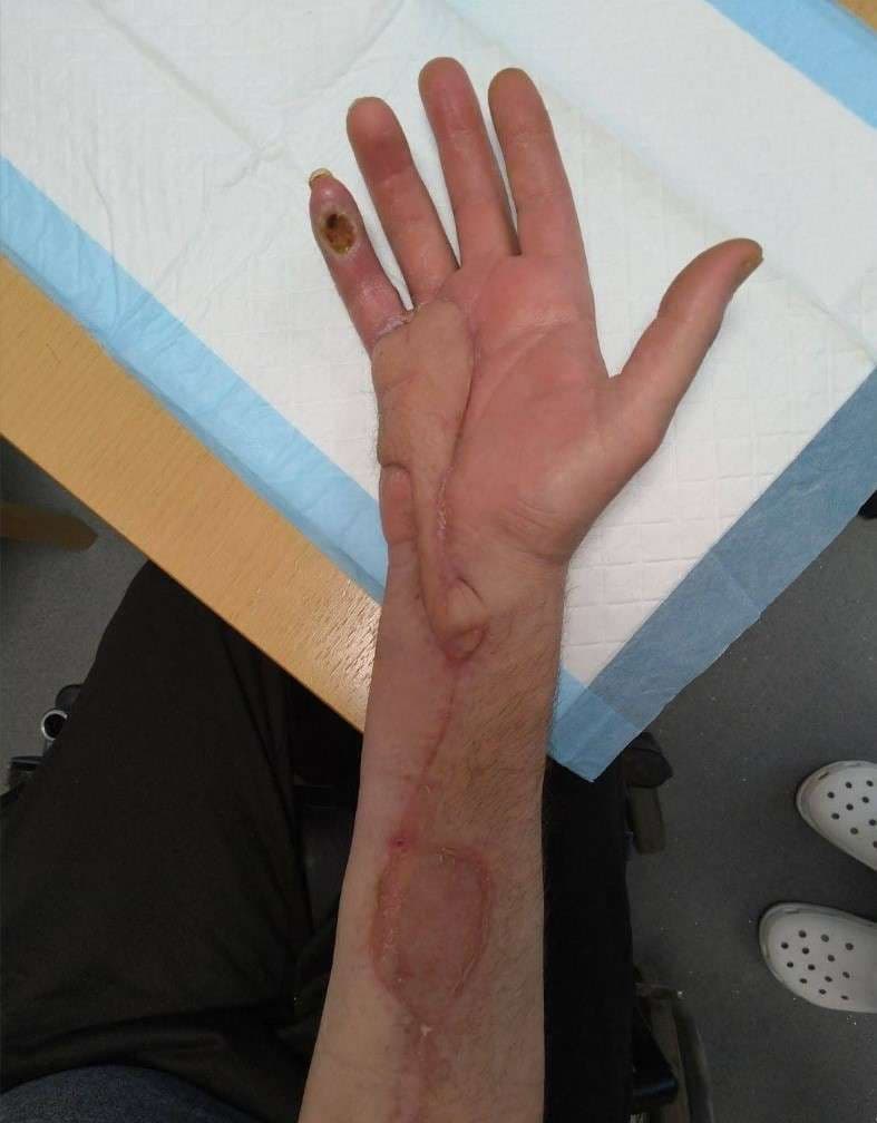
Case 16: Utilizing the reverse radial forearm flap for burn wound management of the hypothenar part of hand
David Salim, MD, Msc
Keywords: Reverse radial forearm flap, Burn wound management, Hand surgery Authors: David Salim, MD and Taiba Alrasheed, MD, FRCSC, Msc. Institution: Department of Plastic- & Breast Surgery , Zealand University Hospital Roskilde, Denmark Abstract A 55-year-old man had sought and was admitted to the emergency department one week after sustaining a superficial second degree burn wound on the hypothenar region of the right hand after accidentally placing it on a hot stove. At first sight, the burn wound seemed quite superficial and conservative healing and bandaging was initially deemed sufficient with the aim of spontaneous healing. One week after the initial assessment in the emergency department, the patient was seen for a routine clinical control of the wound. At this consultation, the patient scored 4/4 positive Kanavel’s signs and flexor tenosynovitis was suspected. Afterwards immediate wound revision of the right hypothenar was conducted. During wound revision, considerable amounts pus was noted and furthermore the musculus abductor digiti minimi was avital and excised. Suitable wound dressing was applied, pus was sent for culture and sensitivity testing and the patient was gives appropriate peroral antibiotics. After numerous wound revisions and debridement, control of the infection at the orthopedic surgery department was achieved. The patient was then referred to the plastic surgery department for planning of soft tissue coverage with a reverse radial forearm flap. Patient medical history The patient was known to have substantial unregulated diabetes mellitus type two, but no other known medical comorbidities. Before and After Patient examination The patient was seen in an outpatient setting in the plastic surgery department for planning of surgery. The defect was located to the whole hypothenar region of the right hand. Substantial soft tissue loss was noted and the 3-5th flexor tendons were exposed. Pre-operative considerations A reverse radial forearm
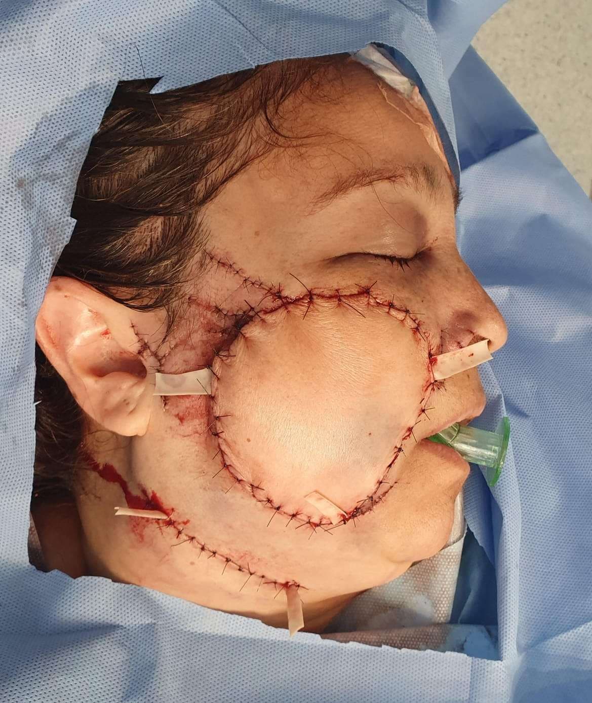
Case 15: Closure of a complex defect after a dog bite wound of the middle third of the face
Dr. Guillermo Alvarez-Sanchez, Ignacio Lugo-Beltran
Keywords: Cheek reconstruction, face, cheek, forearm, free flap, Neurorrhaphy , radial forearm flap Authors: Dr. Guillermo Alvarez-Sanchez, MD ; Ignacio Lugo-Beltran, MD, Chief, Dr. Haro-Cruz Jorge Said, MD. Institution: Plastic and Reconstructive Surgery Division of Microsurgery, Hospital Angeles Metropolitano, Cuauhtémoc, Mexico. Abstract A 52 year old female who presents to the emergency room after receiving a dog bite in her right face, the defect is managed with a free radial forearm flap. Patient medical history 52 year old female, without past medical history, presented to the emergency room, referring she was attacked by her dog at her house, suffering a bite on her right cheek, with bleeding and loss of skin Before and After 1 Before photo - Arrival at the ER Facial asymmetry, with a complex wound on the right cheek, with skin loss, a defect approximately 10 x 9 cm, irregular borders, with muscle exposure, visualizing blood vessels and section of the buccal and zygomatic terminal nerve branches, active mild bleeding. Intact parotid gland and Stensen duct. Laceration of the zygomatic ligament. Difficulty mobilizing of the zygomatic and buccal branches muscle groups. Patient examination Facial asymmetry, with a complex wound on the right cheek, with skin loss, a defect approximately 10 x 9 cm, irregular borders, with muscle exposure, visualizing blood vessels and section of the buccal and zygomatic terminal nerve branches, active mild bleeding. Intact parotid gland and Stensen duct. Laceration of the zygomatic ligament. Difficulty mobilizing of the zygomatic and buccal branches muscle groups. Pre-operative considerations Given the circumstance of tissue loss, active bleeding and section of the nerve branches, also being a contaminated wound, we decided to go to the operating room immediately Due to tissue loss dimensions, we planned a radial free flap for tissue coverage 2 Debridement and assessing damages we cleansed, remodeled the irregular borders and clos