Case 07 2026: Reconstruction of a Complex Epigastric Abdominal Wall Defect Following Wide Excision of Giant Fibrosarcoma Using Prolene mesh and Modified Keystone Flap in a Resource-Limited Setting: A Case Report”
Abdirahman Abdifatah Mohamed, Numan Omar Ibrahim, Rose Alenyo, Kalanzi Edris
Published · May 28, 2026
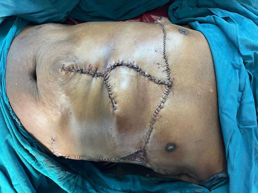
Keywords: fibrosarcoma; Soft tissue sarcoma; Abdominal wall tumor; Wide local excision; Keystone flap; Uganda Authors: Abdirahman Abdifatah Mohamed, MD Final Year PRS Trainee 1, Numan Omar Ibrahim, MD PRS Trainee 2, Rose Alenyo, MD Consultant Plastic Surgeon 3, Kalanzi Edris, MD Senior Consultant Plastic Surgeon 4. Abstract Fibrosarcoma is a rare soft tissue sarcoma with aggressive local infiltration and high recurrence risk. We present a 71-year-old Ugandan man with a giant epigastric fibrosarcoma causing a complex abdominal wall defect. Imaging showed invasion of subcutaneous tissue and rectus muscle without metastasis. The patient underwent wide local excision with 5 cm margins, followed by reconstruction using Prolene mesh and a modified keystone flap with skin grafting. Recovery was uneventful, demonstrating effective surgical and reconstructive management in a resource-limited setting. Patient medical history a 71-year-old male who presented with a two-year history of a progressively enlarging anterior abdominal wall mass. The lesion initially appeared as a small, firm nodule and gradually increased in size, eventually becoming a large, fungating tumor associated with ulceration, necrosis, and intermittent bleeding. There was no history of distant symptoms suggestive of metastasis. Imaging confirmed a locally invasive epigastric mass involving the subcutaneous tissue and rectus abdominis muscle, without visceral or distant spread. Before and After Patient examination On physical examination, the patient had a large, exophytic, fungating mass located in the supraumbilical (epigastric) region of the anterior abdominal wall. The lesion was deeply invasive with irregular margins and was associated with ulceration, areas of necrosis, and active bleeding. The overlying skin was stretched, tense, and partially ulcerated. The mass appeared fixed to the underlying abdominal wall musculature, suggesting muscle involvement. There were no clinical signs of peritoneal invo
References
- Hadley GP. Management of soft tissue sarcomas in sub Saharan Africa. S Afr J Surg.
- Lukande RL Wabinga HR Tumwine LK. Soft tissue sarcomas in Uganda a histopathologic appraisal