Case 14 2026: Nail removal and punch biopsy - Subungual Melanoma
Linnea Kristensen Ejiofor
Published · May 28, 2026
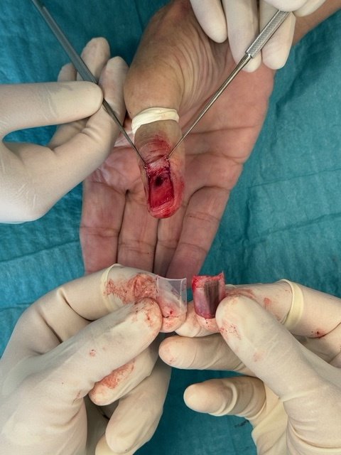
Keywords: Nail removal, nail bed biopsy, nail punch biopsy, Subungual Melanoma Authors: Linnea Kristensen Ejiofor, MD, Department of Plastic Surgery and Burns Treatment, Copenhagen University Hospital, Rigshospitalet, and Magnus Balslev Avnstorp, Specialist plastic surgeon, Zealand University Hospital, Roskilde. Abstract Nail changes can reflect a wide spectrum of benign and malignant conditions, making accurate clinical evaluation essential. This case report reviews key background knowledge on common nail alterations and their differential diagnoses. It aims to support clinicians in recognizing when a nail biopsy is indicated and outlines practical considerations for performing the procedure. In addition, the report describes essential steps in the biopsy technique and provides guidance on appropriate postoperative management and follow-up. Patient medical history A 72-year-old man was referred to the department of plastic surgery and breast surgery at Zealands University Hospital, Roskilde, on suspicion of malignant melanoma under the nail of his right thumb. Before and After Patient examination The patient presented with hutchinson’s sign, longitudinal melanonychia and no swollen lymphnodes. He had no first-degree relatives with melanoma, no prior history of melanoma (invasive or in situ) and no prior history of other skin cancers. Pre-operative considerations Background on Subungual Melanoma (SM) SM is a rare but serious malignancy arising from the nail matrix. Early diagnosis is crucial for treatment and prognosis. Nail changes are often benign; however, malignancy must always be considered in cases of pigmented nail lesions, particularly longitudinal brown or black streaks (longitudinal melanonychia). Biopsy is required when diagnostic uncertainty persists after clinical and dermoscopic evaluation. The incidence of SM is not directly associated with ethnicity in terms of increased risk; however, SM accounts for up to 33% of all melanoma cases in individuals wi
References
- Haneke E. Anatomy of the nail unit and the nail biopsy. Semin Cutan Med Surg. 2015;34(2):95-100
- ollina U, Nenoff P, Haroske G, Haenssle HA. The diagnosis and treatment of nail disorders. Dtsch Arzteblatt Int. 2016;113(29-30):509-18.
- Singal A, Bisherwal K. Melanonychia: etiology, diagnosis, and treatment. Indian Dermatol Online J. 2020;11(1):1-11
- Perrin C, Michiels JF, Boyer J, Ambrosetti D. Melanocytes pattern in the normal nail, with special reference to nail bed melanocytes. Am J Dermatopathol. 2018;40(3):180-1
- Agner T, Jensen AN, Sachs C. Negleforandringer, 2020. https://www.sundhed.dk/sundhedsfaglig/laegehaandbogen/hud/symptomer-og-tegn/negleforandringer/ (22. jun 2022).
- Agner T, Jensen AN, Sachs C. Paronychion, 2021. https://www.sundhed.dk/sundhedsfaglig/laegehaandbogen/hud/tilstande-. 11
- Littleton TW, Murray PM, Baratz ME. Subungual melanoma. Orthop Clin North Am. 2019;50(3):357-366
- Jellinek N. Nail matrix biopsy of longitudinal melanonychia: diagnostic algorithm including the matrix shave biopsy. J Am Acad Dermatol. 2007;56(5):803-10
- Moossavi M, Scher RK. Complications of nail surgery: a review of the literature. Dermatol Surg. 2001;27(3):225-8.