
Case 8 (2024): Right ala nasi reconstruction with a superior based nasolabial flap
David Salim, Roskilde, SUH, Denmark
Abstract
58-year-old female, otherwise healthy, presented to the Department of Plastic Surgery in March of 2024 with a 2 mm punch biopsy verified case of infiltrative basocellular carcinoma. The lesion was located to the right ala nasi subunit of the nose. In December 2023 a primary 2 mm punch biopsy was performed on the same lesion showing basophil degeneration. Due to progression in size in the meantime,
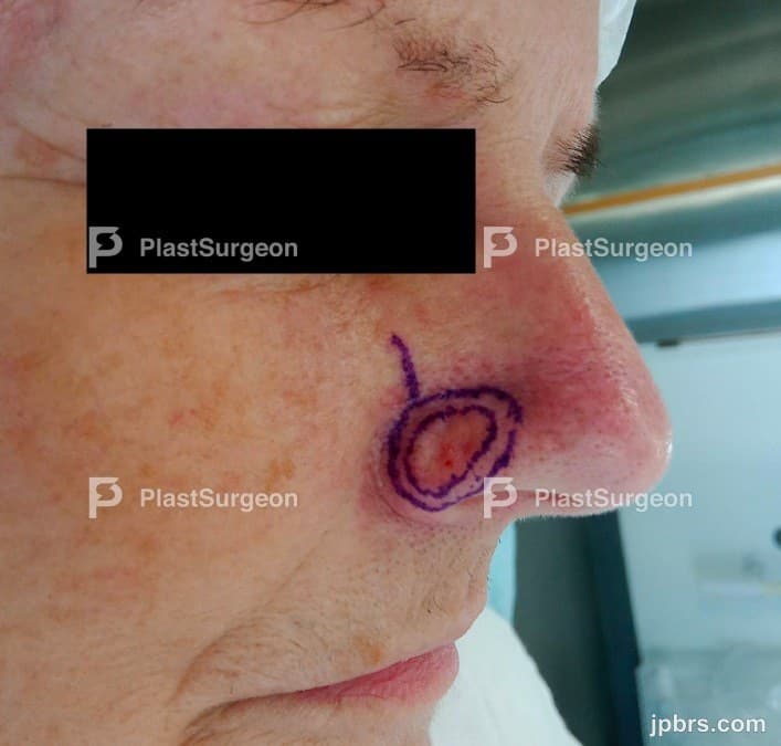

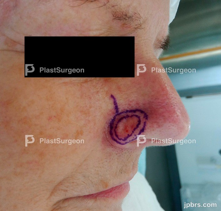 BeforeAfter
58-year-old female, otherwise healthy, presented to the Department of Plastic Surgery in March of 2024 with a 2 mm punch biopsy verified case of infiltrative basocellular carcinoma. The lesion was located to the right ala nasi subunit of the nose. In December 2023 a primary 2 mm punch biopsy was performed on the same lesion showing basophil degeneration. Due to progression in size in the meantime, a new punch biopsy was taken by a private practicing dermatologist, revealing the before mentioned infiltrative basocellular carcinoma.

After applying local anesthesia (1% lidocaine + adrenaline), the tumor was excised with a 3 mm safety margin with an appropriate depth in the underlying soft tissue. Hemostasis was performed. Frozen sections were taken from the borders of the defect at 12, 3, 6, and 9 o’clock and the base of the defect – the sections were sent to fast-track histological examination. Answer was obtained approximately one hour later.
2
Picture of the bottom of the specimen marked superiorly with a safety needle pointing towards the medial canthus of the right eye (pencil marking can be seen on the photo before surgery). Notice the level of excision including the skin, subdermal fatty tissue and not deeper. There is no cartilage in the ala nasi.
3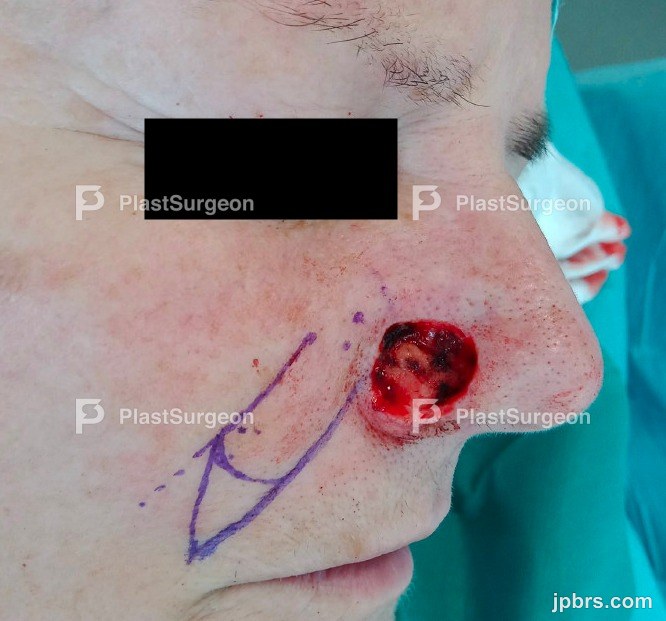
In this picture, the 12 o’clock frozen section is displayed. When taking the sections, it is imperative to take a suitable amount of tissue for analysis. It is a fine balance between not too large of a section and at the same time not too small preventing proper analysis.
4
A superior based nasolabial flap was drawn on the right side – the drawing was based on the dimensions of the defect so it would fit appropriately herein. The width of the flap corresponded the width of the defect. The flap was drawn 2-3 mm longer to avoid traction on the ala nasi. The flap was designed in the nasolabial crease with correction of skin excess in the flap apex.
5
Frozen sections showed no malignancy and the appropriate flap was elevated, prior to this local anesthesia (1% lidocaine + adrenaline) was administered. The flap was dissected at the subcutaneous level and transposed into the defect on the ala nasi – suitable debulking and thinning of the flap was conducted bearing the aesthetic shape of the ala nasi subunit in mind.
6
Donor-site was sutured with 5-0 monofilament and nylon 5-0. Defect-site with nylon 5-0. The ala nasi crease can be seen fairly appropriately reconstructed, mimicking the contralateral side.
7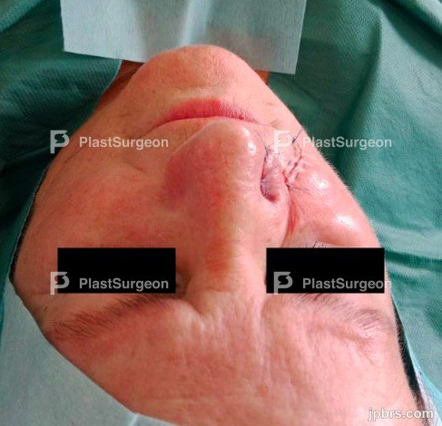
Anterior view after suturing
8Here the reconstructed ala nasi crease can be seen and compared to the contralateral side.

- Thinning of the flap: The flap can be thinned to avoid a too voluminous flap on the ala nasi.
- Vascular supply: understanding the vascular anatomy of the nasolabial flap is imperative. The superior pedicle relies on branches from the facial artery, ensuring proper blood supply to the flap and avoiding necrosis – careful planning might help mitigate this issue, especially the flap pedicle should be thought of.
- Preservation of aesthetic subunits: it is important to pay attention to preserving the aesthetic appearance of the nasal subunit that is to be reconstructed in order to maintain a natural appearance – in the case of the ala nasi subunit, particularly the ala crease and bulkiness of the flap should be considered.
How to cite this article
David Salim, Roskilde, SUH, Denmark. Case 8 (2024): Right ala nasi reconstruction with a superior based nasolabial flap. Journal of Plastic, Breast & Reconstructive Surgery. 2024.
Open access. © 2024 The Author(s). Published by the Journal of Plastic, Breast & Reconstructive Surgery. Copyright to this case is shared between the author(s) and the journal; authors may reuse their own case for education, presentations and social media.
Keep reading
Related cases
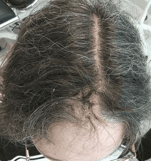
Case 20 2026: Staged Excision and Delayed Primary Closure of a large Nodular Basal Cell Carcinoma of the Anterior Scalp
Mikkel Halborg Sørensen, Nanja Gotland Sundstrup, Christian Lyngsaa Lang
Keywords: Basal cell carcinoma, scalp reconstruction, staged excision, primary closure, scalp biomechanics. Authors: Mikkel Halborg Sørensen, MD; Nanja Gotland Sundstrup, MD; Christian Lyngsaa Lang, MD. Institution: Department of Plastic Surgery and Burns, Rigshospitalet, Copenhagen University Hos-pital, Denmark Abstract A 48-year-old woman was referred with a non-radically excised nodular basal cell carcinoma of the anterior scalp complicated by wound infection and dehiscence. Re-excision with 3 mm margins to the galea was performed under local anesthesia. Given local inflammation and uncertain margin status, delayed reconstruction was planned pending histopathological confirmation. After clear margins were verified, controlled subgaleal undermining allowed tension-reduced primary closure. This staged strategy ensured oncologic safety while preserving hair-bearing scalp and avoiding unnecessary flap reconstruction. Patient medical history A 48-year-old woman was referred after incomplete excision of a nodular basal cell carcinoma (nBCC) of the anterior scalp performed in private plastic surgery practice. Histology demonstrated tumor involvement of the lateral margins, while the deep margin was free of tumor. Postoperatively, wound rupture and infection developed. Microbiology showed sparse growth of Staphylococcus aureus, and antibiotic therapy was initiated. The patient was otherwise non-smoking and healthy, with well-treated asthma, as her only relevant comorbidity. Before and After Patient examination Clinical examination revealed a 22 × 19 mm anterior scalp defect with limited perifocal erythema and minimal purulent discharge. No regional lymphadenopathy was palpable. Pre-operative considerations Initial histopathology demonstrated nodular basal cell carcinoma with focal lateral margin involvement and a clear deep margin, without perineural invasion or other high-risk features. In accordance with current recommendations for low-risk BCC, re-excision with 3 mm c
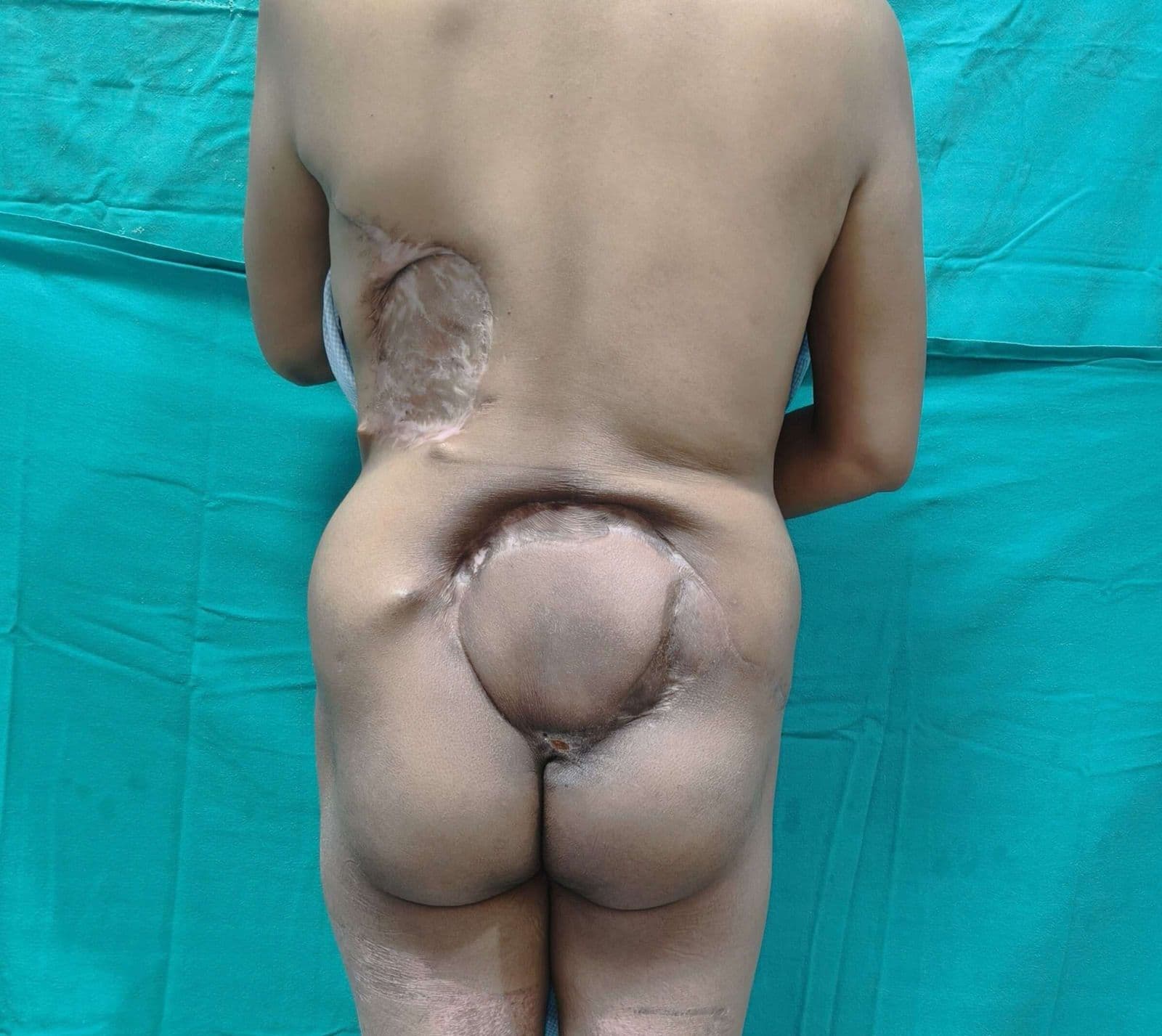
Case 22 2026: Superior Gluteal vessel: A rarely used recipient for free flap reconstruction of lumbosacral defects: a case report.
Tushar Dutta, Arun PS, Swati Sattavan
Keywords: Superior gluteal vessel, free flap reconstruction, lumbosacral defects Authors: 1. Tushar Dutta, Consultant Plastic Surgeon; 2. Arun PS, Consultant Surgical Oncologist; 3. Swati Sattavan, Consultant Surgical Oncologist; Institute: State Cancer Institute, Guwahati, Assam, India Abstract Lumbosacral defects that are too large for local flap reconstruction should be considered for microvascular free tissue transfer. However, lack of suitable recipient vessels in the area makes the task technically difficult. Options such as thoracodorsal and inferior gluteal vessels extended with vein grafts and AV loops have been described. Each option has its own drawbacks. Despite having a short course at its origin, the superior gluteal vessel is a viable alternative. We describe here a case where the vessel was dissected out with adequate length and used as a recipient for free flap reconstruction. Patient medical history The patient was a 41-year-old lady with no known comorbidities. She presented to us with a large lumbosacral mass that had been growing for one year. Before visiting our center, she had been operated twice at other institutions and had recurrences after each occasion. During the last three months the mass had started to grow rapidly and caused ulceration. There was no pain or fever. There was no neurological involvement of lower limbs. Patient had an ECOG status 1. Before and After Patient examination A 17x 22 cm mass was noted encompassing the left part of lumbosacral region and crossing the midline to the opposite side. The superior limit of the lesion was at the L3 vertebra and inferior extent was near the coccyx. Skin ulceration was noted at the center of the mass. On palpation, the mass was warm to touch, hard in consistency and immobile. Skin was fixed and fixity was also noted to the underlying muscle. No tenderness was elicited. Surrounding possible donor sites for flap harvest showed no scars. Pre-operative considerations Preoperative biopsy co
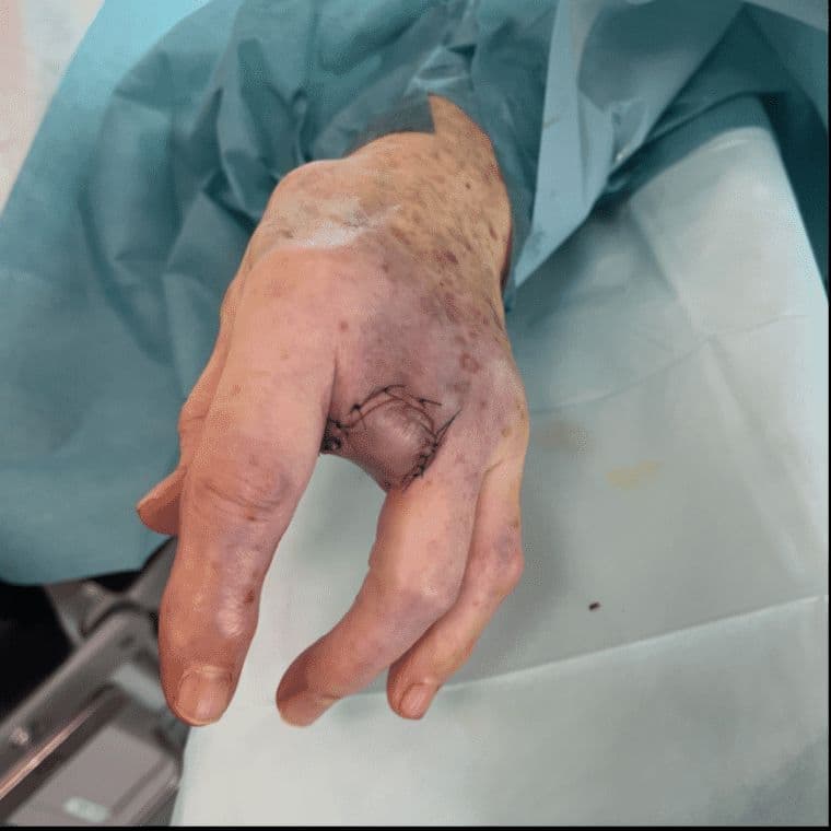
Case 23 2026: “Minimally Invasive Finger Amputation Using a Volar ‘Toilet seat’ Flap for Invasive Squamous Cell Carcinoma”
Claes Hannibal Killerich, Nikolaj Warming
Keywords: Squamous cell carcinoma; finger amputation; volar flap; local reconstruction; traction neurectomy; hand surgery. Authors: Claes Hannibal Killerich, Nikolaj Warming Abstract A 93-year-old woman presented with a chronic non-healing dorsal finger wound initially diagnosed as actinic keratosis. Repeat biopsy revealed invasive squamous cell carcinoma with joint and bone involvement. Due to the extent of invasion, distal amputation of the third finger was required. Reconstruction was performed using a volar “toilet seat” flap under digital nerve block to minimize surgical burden. The procedure achieved clear margins, preserved stump length, and avoided donor-site morbidity. This case highlights the importance of early re-biopsy of non-healing lesions and adapting surgical strategy to patient age, function, and comorbidity. Patient medical history A 93-year-old woman presented with a wound on the dorsal aspect of the proximal phalanx of the left third finger. A primary biopsy revealed actinic keratosis, and the lesion was treated several times with curettage and electrodesiccation by a private dermatologist. Despite repeated treatments under professional supervision, the wound failed to heal.Due to the persistent non-healing nature of the lesion, a repeat biopsy was performed, confirming the diagnosis of squamous cell carcinoma (SCC). The patient was referred to the Department of Plastic Surgery, Aalborg University Hospital, where a primary excision was performed with a 7 mm surgical margin. Histopathological examination demonstrated invasive tumor growth involving both joint and bone. The defect was subsequently reconstructed using a full-thickness skin graft. Before and After Patient examination The left third finger showed a healed full-thickness skin graft on the dorsal aspect of the proximal phalanx, covering the proximal interphalangeal (PIP) joint. There was no visible residual tumor, and no palpable lymphadenopathy in the left cubital fossa or axillary re